Medical imaging made a major leap forward when it moved from 2D images to 3D renderings. But the radiology department's primary product -- the radiology report -- is still based on archaic information that's 2D and text-based. A joint team from the U.S. and Singapore has developed a way to make radiology reports come alive -- in 3D.
In an article published online August 24 in the Journal of Digital Imaging, researchers from the National University of Singapore and Weill Cornell Medical Center in New York City shared how their open-source 3D visualization and reporting tool, called Visual Interpretation with Three-Dimensional Annotations (VITA), can produce 3D visual summaries of imaging studies based on radiological annotations.
These 3D summaries show promise for enhancing communication between radiologists and referring physicians, as well as with patients, according to the team led by Sharmili Roy, PhD, of the National University of Singapore.
When the framework was tested with seven referring physicians, all agreed that the 3D visual summaries improve the clarity of communication between radiologists and physicians. Furthermore, six of the seven agreed that the 3D reports aid patient communication, and six were willing to use the system in their routine clinical practice, according to the researchers.
A missing piece of the puzzle
In current radiological practice, image-based annotations prepared by radiologists during exam interpretation are often not accessible to referring physicians, who have to rely on the text-based reports that summarize the critical radiological findings, Roy told AuntMinnie.com.
"These reports lack the 3D contextual information of the observations and are often difficult for patients to comprehend," she said.
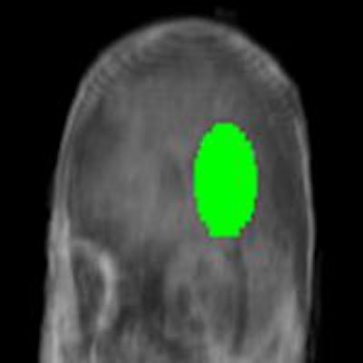
As a result, Roy and colleagues Dr. George Shih and Michael Brown, PhD, sought to utilize the images and the annotations to compute an animated, 3D visual summary of the exam findings that embeds the radiological observations in a rotating volumetric rendering of the anatomy. The 3D reports are nondiagnostic and are distinct from 3D renderings that a radiologist might use to interpret an image.
Under the framework developed by the group, annotations are clearly highlighted as the volume spins, providing a comprehensive summary of the important clinical content, according to Roy.
"The 3D nature of the report facilitates patient's comprehension of the pathology and thereby enhances physician-patient communication," she said.
 A text-based radiology report versus the VITA 3D visual report. VITA 3D visual reports place radiological observations into a 3D volumetric context designed to make radiology reports more comprehensible to patients and enhance physician-patient communication. Image courtesy of Sharmili Roy.
VITA is currently compatible with the open-source ClearCanvas PACS workstation software, but it can also be used with any PACS workstation software that employs structured annotations such as Extensible Markup Language (XML) or the Annotation and Image Markup (AIM) standard, for example.
No user interaction is required for VITA, which can run in the background and automatically generate and archive the 3D summary images, according to the authors. On average, VITA can produce a visual summary for an exam consisting of 300 images in less than one minute on a computer using an Intel Core i5 2.4-GHz processor with 3 GB of RAM and an Nvidia GeForce GT 330-M graphics card.
User satisfaction study
To assess whether the visual summary generated by the framework enhances radiologist-physician communication and is usable in day-to-day clinical practice, the researchers performed a user satisfaction study with seven physicians in Singapore.
All physicians felt that the 3D visual summaries improved the clarity of their communication with radiologists, while six of the seven agreed that the 3D visual summaries aided patient communication. One physician was neutral on the issue.
Six of the physicians indicated they were willing to use the system in their routine clinical practice.
The physicians also offered a number of positive comments, such as "it is a new brilliant concept for patient understanding," and "this is an excellent intervention which helps in better collaboration between [the] physician and radiologist," Roy said.
The one physician who was neutral on using the 3D visual summaries commented that "3D rendering does not add additional information for clinician. It looks nice for the layperson, i.e., patient, but clinical use is very limited," according to Roy.
New VITA version
The latest version of VITA automatically clusters multiple annotations belonging to one volume and automatically segments the volume represented by these annotations.
"The visual report highlights the 3D segmented anatomy instead of highlighting individual annotations," she said. "Thus, we are able to automatically extract 3D information from 2D radiological annotations."
Regarding future plans, the researchers hope to extend VITA to allow visualization of pathology progression by computing visual summaries from exams taken at various points of the treatment.
"The goal is to automatically compute and highlight enhancing or retreating edges in tumors," Roy said.
Patient empowerment
In light of the recent trend toward patient empowerment, the 3D images of the VITA visual report are well-suited for patient-radiologist communication, Roy said.
"The 3D nature of the images in the visual report and the embedded annotations therein can greatly enhance patients' comprehension of the pathology," she said. "Patients can be given visual reports in video formats like MPEG-4 or Flash, so they can view the important findings noted by the radiologist on the imaging study without having to access a PACS workstation."