A half dose of the MRI contrast agent gadobenate dimeglumine provides just as much relevant diagnostic information as a full dose in 3-tesla imaging of patients with early rheumatoid arthritis, according to a study published in the July issue of Radiology.
The researchers found that a contrast dose of 0.05 mmol/kg is enough to detect synovitis, or inflammation of the synovial membrane, which can help diagnose rheumatoid arthritis.
"Our results show some promise for further reduction of the dose of contrast media without the loss of any diagnostic information in patients with rheumatoid arthritis at 3-tesla MR imaging," wrote lead author Dr. Claudia Schueller-Weidekamm and colleagues from the Medical University of Vienna and Vienna General Hospital (Radiology, Vol. 268:1, pp. 161-169).
Earlier diagnosis
The early diagnosis of rheumatoid arthritis greatly improves patient outcomes because therapeutic treatment can begin sooner, the authors wrote. In addition, the use of gadolinium-based contrast agents has become a concern in recent years due to their association with nephrogenic systemic fibrosis (NSF) in patients with impaired renal function.
"The aim of this study was to investigate whether it was possible to halve the standard amount of contrast medium (0.1 mmol/kg) without any loss of diagnostic information due to the higher relaxivity of gadobenate dimeglumine," they wrote.
Between August 2009 and September 2011, the researchers enrolled 57 consecutive patients with early rheumatoid arthritis. All patients underwent contrast-enhanced MRI of their dominant hand (39 right-handed and 18 left-handed participants) at the University Hospital of Vienna.
MRI scans were conducted on a 3-tesla system (Achieva, Philips Healthcare) with patients in a prone position. The dominant hand was placed above their head and a sensitivity encoding (SENSE) Flex-M radiofrequency coil was positioned over the hand. A saline syringe was fixed on the fifth finger to help with postprocessing, according to the authors.
Gadobenate dimeglumine (MultiHance, Bracco) was measured in inflamed synovial tissue at half dose (0.05 mmol/kg of body weight) and at full dose (0.1 mmol/kg) using T1-weighted MRI sequences with fat saturation. Each contrast injection was followed by a 20-mL saline flush.
Two readers who were blinded to the patient data separately analyzed all images, with the half-dose and full-dose MR images mixed before evaluation. Their analysis included quantitative measurements of signal intensity in the inflamed synovial tissue, signal-to-noise ratio, and contrast-to-noise ratio.
Signal intensity
The calculations revealed a high degree of correlation for signal intensity of synovial enhancement at the half dose and full dose of contrast. The mean signal intensity of synovial enhancement was 914.35 (± 251.1) with a half dose of contrast, compared with 1,022.6 (± 244.5) for a full dose of contrast. In the saline syringe, the average signal intensity was 642.82 (± 196.2) at half dose and 554.12 (± 162.5) at full dose.
The researchers did find, however, that the full dose of gadobenate dimeglumine had significantly better signal-to-noise and contrast-to-noise ratios than the half dose. The mean signal-to-noise ratio was 42.91 for the half dose, compared with 54.22 for the full dose, while the mean contrast-to-noise ratio was 12.81 for the half dose, compared with 24.78 for the full dose.
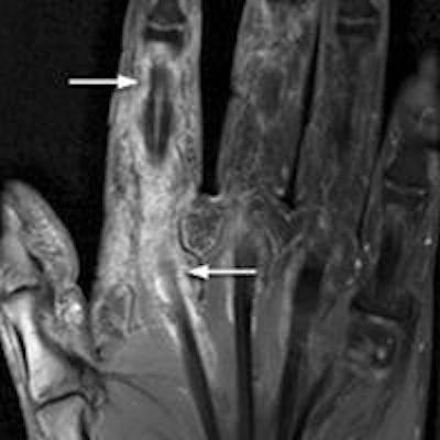
 T1-weighted, fat-saturated, postcontrast 3-tesla MRI shows a half dose (a) and a full dose (b) of gadobenate dimeglumine in a 47-year-old man with early rheumatoid arthritis. The tendon sheath of the flexor tendons illustrates increased synovial enhancement, with tenosynovitis of the second flexor tendon scored as grade 3 (arrows) at both half dose and full dose. Images courtesy of Radiology.
Despite the disparity in contrast-to-noise and signal-to-noise ratios between the dosages, the diagnostic quality of the images was not affected, according to the authors. No region of synovial inflammation seen on full-dose contrast-enhanced MRI was missed at half dose.
"These results demonstrate that no diagnostic information that would be relevant for diagnosing and staging rheumatoid arthritis, therapy planning, therapy monitoring, or follow-up examinations was missing from half-dose images," the authors wrote.
"The image quality of all anatomic structures and pathologies, except for the assessment of cartilage, was rated as excellent," they wrote, adding that the lower result for cartilage "might be explained by the suboptimal resolution for minor cartilage defects at 3-tesla [MRI]."
In conclusion, Schueller-Weidekamm and colleagues confirmed the feasibility of using half the standard dose (0.05 mmol/kg) of gadobenate dimeglumine, with no loss of relevant diagnostic information at 3-tesla MRI, for patients with early rheumatoid arthritis.