There's still a lot to learn about how bones interact in joints during movement, but 4D musculoskeletal (MSK) CT can help close that knowledge gap, paving the way for new understanding of joint disorders and how to treat them, according to researchers from Monash Health Centre in Melbourne, Australia.
The method, which leverages the wide field-of-view of a 320-detector-row MDCT scanner for a volumetric acquisition of patient joint movement, may also spark newer classifications of injuries thanks to its capability to describe previously undefined subtle changes in joint architecture during motion, the researchers told AuntMinnie.com.
"[Musculoskeletal] problems when they occur are quite often complex in nature, and we see this technique as becoming a routine tool for the evaluation of joint pathology, especially where coexisting conditions exist or where there are concerns over the outcomes of more aggressive treatments," said Dr. John Troupis, unit head of musculoskeletal imaging and co-unit head of cardiac CT at Monash Health.
He presented the research at the recent American Roentgen Ray Society (ARRS) annual meeting in Washington, DC. The Monash team is working with clinical application scientists from Toshiba Medical Systems to develop the hardware and software involved with the method.
The unknown
Much remains unknown about the interactions of the joints during motion. While pain and movement restriction are indicators that something is wrong, in an aging population a number of coexisting diseases and pathology may be involved in any one patient or joint, Troupis said.
"Dynamic CT scanning provides the opportunity to look at the complicated interactions between the bones and soft tissue as the joint moves, providing the opportunity to better isolate the cause of the problem," he said.
  Normal wrist motion in flexion and extension. Note that the lunate tracks the movement of the hand while remaining seated in the radius. All images and movies courtesy of Dr. John Troupis and Dr. Benjamin Amis.
|
Thanks to recent technology developments such as 320-detector-row CT, it's now possible to perform a 4D isotropic CT evaluation on joints, Troupis said.
"We believe that while static images are great for looking at something like a fracture, the ability to see how the bones of the wrist interact during complex motions of the hand are vital if we are to fully understand the mechanisms of injuries," he said. "These sorts of dynamic procedures have been tried in the past with CT, but it is only with the technology available now that we are able to perform a volumetric acquisition where we can acquire a thick slab of data during motion."
A wide field-of-view
The 4D MSK imaging technique makes use of the wide field-of-view possible with the 320-detector-row array. The institution's Aquilion One CT scanner (Toshiba) offers 16 cm of z-axis coverage for acquiring images, "which is great for most joints like the wrist, ankle, shoulder, and knee, but we also need to have the patient perform the required movement in time with the 350-msec rotation speed of the scanner," Troupis said.
Patients must therefore be trained how to move while in the CT scanner. If the patient moves too quickly, the cine acquisition will be blurred, and if he or she moves too slowly, a truly dynamic movement of the joint won't be acquired, he said.
After patients successfully perform a series of practice runs to get the timing and to ensure the full range of motion, they are positioned in the CT aperture, he said. A CT volume is then acquired continuously for one or two arcs of movement
|
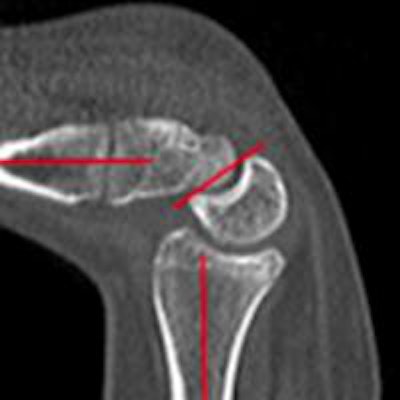
Abnormal wrist in motion. The lunate lags behind the movement of the hand until it snaps or clicks into line when just in flexion. The seating of the lunate in the radius is also affected.
|
"We then break those data up into time phases -- about six per tube rotation -- and produce a cine loop or movie of the motion of the patient," he said. "We can produce a render of the soft tissue and of the bone, but what we can also do is isolate particular bones and perform cine [multiplanar reformats (MPRs)] isolated to that one particular area."
Furthermore, they can have patients recreate the motion that produces the "clicking" and/or their pain, and compare that with either their other "good" side, or with a bank of normal images the researchers are building, Troupis said.
"4D MSK CT is a new technique, and we are constantly learning about what is normal in terms of how joints move as represented by a dynamic CT acquisition," Troupis said. "We have found collaboration with orthopedic surgeons to be extremely valuable as we develop the technique, and they, too, are excited by the opportunities that the technique has in terms of their management of the patient."
In addition, the method has shown that the classification of some pathologies may need to be reviewed due to the new knowledge being gained, according to the researchers. For example, the group used 4D CT to identify trigger lunate syndrome (see image below). A case report on this syndrome will soon be published in the Journal of Computer Assisted Tomography, they noted.
 These images show the complex interaction of the lunate of the wrist in midcarpal instability. Points of extreme flexion and extension and the midpoint of that motion are shown in a normal wrist (top images) and abnormal wrist (bottom images). This trigger-lunate motion abnormality, featuring abrupt cessation and recommencement of flexion/extension, had not been previously recognized radiographically as a feature of midcarpal instability, according to the researchers.
Future steps
In the next phase of development, the researchers would like to be able to produce a 4D musculoskeletal series on a patient's torso at a radiation dose comparable to that of a traditional x-ray series.
"If we could do that, we could certainly start to routinely evaluate the clinically complicated hip, which is a joint commonly afflicted in adolescents," Troupis said. "We are working all the time on improving our technique so that we are continuing to produce useful image sequences for our referrers, and using the lowest dose possible for our patients."
The researchers are also working with manufacturers such as Toshiba to further develop software and hardware to enhance both image quality and the dose profile of the procedure, he said. They are also considering working with other clinical centers for a multinational clinical trial to define normal joint interactions in motion as well as abnormal patterns of motion.
"The future of 4D MSK CT imaging is an exciting one to be sure," Troupis said.