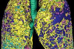
A new algorithm can automatically and accurately segment lung parenchyma on thoracic CT images, potentially improving the performance of lung computer-aided detection (CAD) software, according to researchers from Northeastern University in Shenyang, China.
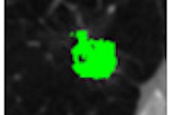
The team's algorithm produced greater than 95% segmentation accuracy for lung parenchyma and 100% sensitivity for including and segmenting the challenging juxtapleural nodules that adhere to the lung wall.
"The algorithm can achieve good results for lung parenchyma segmentation and repairing in various cases that nodules/tumors adhere to the lung wall," said lead author Ying Wei, PhD.
The researchers shared their findings in an article published online October 3 in the Journal of Digital Imaging.
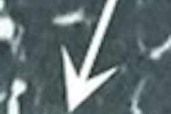
The segmentation of lung parenchyma is an important early step in the CAD process, but current segmentation techniques suffer from a number of drawbacks, including difficulty in handling juxtapleural nodules. These nodules, which adhere to the lung wall, are often mistaken by CAD methods as fat to be excluded, and the corresponding region in the segmented lung parenchyma becomes a sunken area in the lung wall, Wei said.
Because these nodules/tumors are important in diagnosis, researchers have been trying to address this problem with new techniques, including the commonly used "rolling-ball" method. That approach can't accurately judge the size of the defect, however, Wei said.
As a result, the researchers sought to employ a new method for lung parenchyma segmentation and repairing, involving techniques such as the optimal iterative threshold, 3D connectivity labeling, and 3D region growing for the initial segmentation of the lung parenchyma, followed by improved chain code and Bresenham algorithms to repair the lung parenchyma, Wei said.
The researchers tested the segmentations from the automated algorithms by comparing them with manual segmentations that were traced by three experienced chest radiologists on all slices from the top to bottom parts of the lung. These manual segmentation results were judged by a panel and used as the gold standard for the purposes of the study.
Wei and colleagues then compared the performance of their proposed method and the rolling-ball method on 97 lung nodule thoracic CT scans and 25 juxtapleural nodule scans.
The method yielded 95.2% segmentation accuracy for lung parenchyma, compared with 88.8% for the rolling-ball method. It also produced 100% sensitivity for juxtapleural nodule inclusion and segmentation, and accuracy of 98.6% for juxtapleural nodule regions. Average segmentation time was 0.67 seconds per frame.
"The comparison results show that our algorithm can better adapt various cases of the lung parenchyma segmentation than the rolling-ball method and achieve satisfactory segmentation results," the authors wrote. "From these experimental results, we can see that stable and satisfactory segmentation lung parenchyma results can be obtained automatically by the algorithm proposed in this paper."
The researchers concluded that their proposed method can meet the requirements of a lung nodule CAD system, and provide high-quality preliminary data for sequential processing.
"The robustness of this approach in terms of other types of lung disease such as mesothelioma and emphysema, etc., remains to be verified further," they wrote.