Fractional flow reserve CT (FFR-CT) in patients with significant stenosis can distinguish those with coronary artery disease (CAD) who need revascularization from those who don't, concluded a study presented on Sunday at the European Society of Cardiology (ESC) meeting and published concurrently in the Journal of the American Medical Association.
That said, the novel FFR-CT test didn't quite live up to the sensitivity goals of the study team, achieving only a modest diagnostic accuracy of 74% for detecting flow-limiting lesions in patients with significant stenosis undergoing coronary CT angiography (CCTA). But the study's accuracy result, tied irrevocably to disease prevalence, is less important than robust area under the curve (AUC) results showing FFR-CT's ability to reliably discriminate ischemic from nonischemic stenoses, lead author Dr. James Min, from Cedars-Sinai Heart Institute, told AuntMinnie.com in an interview.
Overall, FFR-CT went a long way toward increasing diagnostic confidence and answering critical questions about the need for invasive intervention, Min said. He and colleagues from 16 other centers and four other countries participated in the Fractional Flow Reserve by Anatomic Computed Tomographic Angiography (DeFACTO) study.
"Use of noninvasive FFR-CT plus CT among stable patients with suspected or known CAD was associated with improved diagnostic accuracy and discrimination versus CT alone for the diagnosis of hemodynamically significant CAD," with invasive fractional flow measurement and invasive coronary angiography serving as the reference standard, the study team wrote in JAMA (August 26, 2012).
Driven by improvements in sensitivity and specificity, FFR-CT combined with CCTA "improved per-patient diagnostic accuracy versus CT alone," and showed that FFR-CT "can impart considerable discriminatory power" to find and rule out ischemia in patients with suspected coronary artery disease, the group wrote.
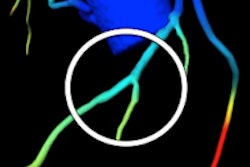
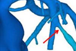
According to Min, FFR-CT's key advantage over tests such as SPECT, perfusion CT, and other functional exams is its ability to determine exactly which coronary artery stenoses are causing ischemia and which aren't. Using FFR-CT, "you can pinpoint the lesion that's causing ischemia -- global perfusion doesn't do that," he said. "It's selectable on every point in the coronary vascular bed, and if you revascularize the patient, you can then go back [with FFR-CT] and look again."
CCTA can't pinpoint cause of ischemia
CCTA results correlate very favorably with invasive coronary angiography for measuring stenosis severity, but CCTA alone can't determine the hemodynamic significance of a coronary artery stenosis -- to the point where even if significant stenosis (≥ 50%) identified at CTA is confirmed using invasive angiography, fewer than half of the lesions actually cause ischemia, Min said.
"If you look at FAME [Fractional Flow Reserve Versus Angiography for Multivessel Evaluation; New England Journal of Medicine, January 15, 2009, Vol. 360:3, pp. 213-224], 20% of the patients don't have severe stenoses and they still have ischemia," Min said. "We're sending home a bunch of patients who feel bad, and we're saying they're OK, but they're not."
On the other hand, as many as 20% of patients with severe stenoses at CCTA turn out not to have ischemia, and these patients risk being referred for invasive coronary angiography when they don't need it, Min said. The specter of unnecessary revascularizations is bolstered by recent randomized study results showing no survival benefit in patients undergoing angiographically based coronary revascularization, the authors wrote in JAMA.
FFR-CT consists of a series of complex flow calculations that represent the noninvasive version of angiography-based FFR measurements. The DeFACTO study aimed to test the accuracy of FFR-CT calculations for diagnosing hemodynamically significant CAD, as defined by an invasive FFR reference standard, in patients with suspected coronary artery disease who were referred for nonemergent invasive angiography.
All patients -- 252 adults with suspected CAD who underwent invasive angiography within 60 days of CCTA -- underwent four tests: CCTA, invasive angiography, FFR, and FFR-CT, all read by independent core laboratories blinded to the results. Three-fourths of patients had experienced angina within the past month. The researchers compared the accuracy of FFR-CT and CTA for diagnosis of ischemia versus an invasive FFR reference standard. They defined a FFR or FFR-CT measurement of 0.80 or less as ischemia, and anatomically obstructive CAD was defined as 50% or greater stenosis at both CCTA and invasive angiography.
Among 615 vessels interrogated for the study, 271 had less than 30% stenosis and 101 had at least 90% stenosis, the study team reported. In all, 137 (54.4%) of the study participants had an abnormal FFR score. FFR-CT improved the prediction of stenosis at invasive angiography at both the 50% and 80% stenosis levels.
Diagnostic performance of FFR-CT for stenosis
|
||||||||||||||||||||||||||||||||||
The diagnostic accuracy of FFR-CT with CTA was 73%, compared with a diagnostic accuracy of 64% for CT alone.
Sensitivity and specificity were moderate, but they aren't the best measure of FFR-CT performance because these measures are affected by disease prevalence, according to Min. The improvement in AUC with the use of FFR-CT is the more important measure, he said.
When FFR-CT alone was compared with CT alone for stenosis of at least 50%, FFR-CT showed better discrimination (AUC = 0.81, 95% confidence interval: 0.75-0.86) than CT (AUC = 0.68, 95% confidence interval: 0.62-0.74, p < 0.001).
"The AUC ends up being the most telling thing because it's immune to disease prevalence," Min said. "The area under the curve either changes or doesn't, but if it does change it's irrespective of how prevalent disease is. It's hard to move AUC, so, for example, if you go from 0.67 to 0.68 and it's statistically significant, that's a great thing. And in this study it went from 0.68 to 0.81, which is a huge difference."
As a result, FFR-CT improved diagnostic accuracy versus CT alone for diagnosing ischemia, even though the study did not satisfy its previously stated primary end point of diagnostic accuracy of greater than 70%, the authors explained. The technique also frequently misclassifies risk categories.
"Despite its superiority to CT alone, the diagnostic specificity and positive predictive value of FFR-CT for ischemia detection remained low, suggesting that while false-positive studies would be reduced with this approach, a substantial rate would remain," the authors wrote. "In this regard, universal application of FFR-CT to guide invasive assessment may result in referral of a non-negligible number of patients without ischemia, and future studies will be needed to determine the potential clinicoeconomic effectiveness of FFR-CT."
But overall, FFR-CT's diagnostic capabilities can encourage a greater sense of diagnostic certainty that patients scanned with CT will not have ischemia overlooked, giving clinicians more confidence that they can skip invasive angiography in patients with negative FFR-CT results, according to the authors.
More work to do
In an editorial accompanying the study, Dr. Manesh Patel, from Duke University Medical Center, agreed that further research is needed to determine the practical utility of the promising FFR-CT technique.
Future studies "should be aimed at diagnostic strategies involving patients with varying pretest risks, thereby providing information on the incremental benefit from the test," Patel wrote. Also needed are comparison tests beyond invasive angiography, although invasive FFR may be a valid test for comparison.
Resource utilization and clinical outcomes also should be reported, he wrote. Ideally, the test would move to a local site rather than a core lab.
At its core, Min said, no other test has FFR-CT's ability to pinpoint the precise artery causing the stenosis.
"[Looking] at the data for myocardial perfusion, for example, if you have a perfusion defect in the anterior distribution, it doesn't necessarily correlate to a [left anterior descending] lesion," he said. "It's good on a per-patient level, but it's not robust enough to geographically locate exactly what stenosis is causing the ischemia. So in this regard, I think the technology has the potential benefit of identifying a specific coronary artery lesion that causes the ischemia, and you can revascularize that lesion as opposed to ones that are highly stenotic but don't necessary cause ischemia."
FFR-CT's performance characteristics suggest a low rate of false negatives for identifying ischemia-causing intermediate lesions, with little effect on the false-positive rate, Patel wrote.
"In this regard, the use of FFR-CT may significantly advance clinical assessment of patients without conventional measures of anatomic high-grade coronary stenosis, largely by proper identification of a significantly greater proportion of patients with manifest ischemia rather than as a safeguard to further invasive evaluation," he wrote.
|
Study disclosures The study was funded by HeartFlow, which had no control over the study design or data, according to author disclosures in JAMA. |