A Canadian study has found that radiologists preferred the image quality of a dual-energy digital radiography (DR) technique over conventional DR in detecting lung lesions. Dual-energy DR produced better soft-tissue delineation thanks to the removal of bony anatomy from images, according to the researchers.
Dual-energy DR is one of a number of advanced technologies that researchers are pursuing in an effort to improve the image quality of DR and make it a more viable alternative to CT for lung imaging. In dual-energy imaging, two exposures are collected with the x-ray system's tube set to different energy levels; the resulting images highlight either bone or soft tissue, or they can be merged into a composite image.
Researchers from the Ontario Cancer Institute at Princess Margaret Hospital in Toronto wanted to determine whether DR could be used for many patients who currently receive CT for lung imaging, according to Dr. Narinder Paul, section chief for cardiothoracic imaging at the hospital. Paul presented the research at the 2008 RSNA meeting in Chicago, and the study is scheduled to be published in an upcoming issue of Academic Radiology.
While CT is highly sensitive, it detects many suspicious lesions that must be worked up, yet turn out to be benign, Paul said. Many patients who are severely ill also cannot be imaged with CT due to the difficulty in transporting them, he said. Meanwhile, conventional radiography, both analog and digital, suffers from the anatomical noise that results from compressing 3D data onto a 2D image.
"One of the reasons why we're seeing a large increase in CT scanning of the chest is that there are pitfalls to chest x-ray, whether it's [computed radiography (CR)] or DR or not, and that's because of anatomical noise -- you're compressing onto a two-dimensional image," Paul told AuntMinnie.com.
To see whether dual-energy imaging could help, the research team developed a prototype dual-energy system by modifying a commercially available digital x-ray chest stand (Carestream Health, Rochester, NY). Their modifications included a filtration wheel on the x-ray tube that enabled the researchers to easily select different kVp energy levels and attenuation. The researchers also adapted the system with a flat-panel DR detector (it normally uses a CR cassette).
Another important modification was a cardiac trigger that enabled the researchers to coordinate the acquisition times of the two dual-energy studies so they occurred during the same period of the cardiac cycle. The researchers used a dual-energy technique in which the two exposures were taken six to eight seconds apart, rather than simultaneously; this method can create registration artifacts unless a technique like the cardiac trigger is used, Paul said. The group also placed BBs on the patients as landmarks to assist with image registration.
The study's patient population consisted of 55 patients who were drawn from a larger study under way at the hospital to assess dual-energy DR. Study subjects were from a population of patients referred for lung biopsy, and many had high-risk backgrounds, such as a history of smoking.
The researchers used different kVp settings for low-energy and high-energy images. For an average patient with 24-cm chest thickness, the standard low-energy setting was 60 kVp with 2.5-mm aluminum total filtration, while the standard high-energy setting was 120 kVp with 4.5-mm aluminum + 0.6-mm silver total filtration. The high-energy filter was selected to harden the beam to improve contrast by reducing spectral overlap between the low-energy and high-energy projections. Radiation dose for the total exam was equivalent to a conventional posteroanterior DR image.
Conventional DR images were acquired immediately after the dual-energy studies on the same system. Researchers used a 120 kVp setting with 1-mm aluminum + 0.2-mm copper filtration. Total radiation dose for both the dual-energy and DR images were equivalent, at about 0.11 mGy for a patient with a 24-cm chest thickness.
Images were processed and registered to produce three images: an image emphasizing soft tissue, one highlighting bony anatomy, and a composite image intended to be nearly identical to a conventional radiograph.
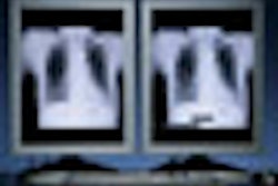
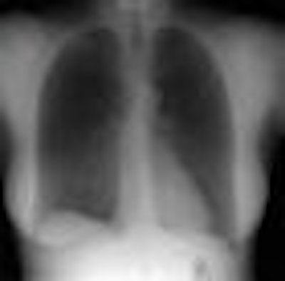
 |
| Image series demonstrates high-energy image (left), low-energy image (center), and soft-tissue image (right) produced by processing both images. |
The researchers said that because of a lack of true-negative cases in the patient cohort, they decided not to use receiver operator characteristics (ROC) analysis and instead focused on reader satisfaction as the measure of diagnostic performance. Five readers were asked to use a nine-point scale to rate each image in terms of their ability to detect and characterize abnormalities. Scores ranged from 1 (very dissatisfied -- abnormalities could be overlooked or mischaracterized) to 9 (very satisfied -- the abnormality is perfectly obvious and is easily characterized).
From the total pool of 275 cases (five readers and 55 cases), the dual-energy image was rated superior to the DR image in 41.5% of cases (114 of 275). In 38.9% of cases (107 of 275), both modalities were rated equally, and in 19.6% of cases (54 of 275) the DR image was rated superior.
The researchers also plotted their results according to the number of cases in which a majority of three readers viewed one modality as superior to the other. By this analysis, 36.4% of cases (20 of 55) were scored with the dual-energy images rated as superior, 36.4% (20 of 55) were rated as equal, and 5.5% (3 of 55) were rated as the DR images being better.
The researchers also found that the preference for dual-energy was more pronounced when readers viewed lesions smaller than 3 cm, and for nodules as opposed to nonsolid lesions. On an anatomical basis, the readers tended to prefer dual-energy images when viewing pathology in the upper lobes of the lung, and expressed less of a preference in the middle or lower lobes.
Regarding future studies, the researchers plan to conduct a second study with 516 patients, using an ROC curve to assess sensitivity and specificity. The team would also like to adapt their technology to a portable dual-energy DR unit that can examine patients who can't come to the radiology department.
Paul believes that one challenge to adopting dual-energy imaging in clinical practice is that the technique requires radiologists to spend more time interrogating images. But dual-energy could be used selectively, in cases where radiologists are trying to resolve a suspicious area.
"We are building tools that will make [dual energy] radiologist-friendly, so you can use the tools or not, depending on what your level of suspicion is," Paul said. "If you read a trauma film, you might want to look at the bone image first."
Could the technology someday make DR a viable lung screening modality? Maybe, according to Paul.
"If we can show that dual-energy imaging can detect the small nodules that we really need to detect, and it doesn't have to detect tiny nodules that are incidental, then it becomes possible to think about using this as a tool for screening," he said.
By Brian Casey
AuntMinnie.com staff writer
January 29, 2009
Related Reading
Adding CsI-based dual-energy imaging doesn't improve lung nodule detection, December 26, 2008
Dual-energy digital x-ray still looking for acceptance, February 22, 2007
Copyright © 2009 AuntMinnie.com