(Radiology Review) New research suggests that cesium iodide (CsI) detector-based dual-exposure dual-energy imaging, when added to chest radiography, does not significantly improve the detection or classification of pulmonary nodules. The researchers chose a prospective, multicenter design to enhance the study's outcome, and sought to determine the clinical value of dual-exposure dual-energy chest radiography with a flat-panel detector.
"Previous studies have suggested potential improvements for the detection of noncalcified as well as calcified lung nodules by cesium iodide (CsI) detector-based dual-exposure dual-energy chest radiography compared to standard chest radiography alone," wrote Dr. Ricarda Röhl and colleagues from the University Clinic Magdeburg, Germany.
However, limitations due to monocentric design and limited patient numbers may have limited the studies' outcome, they noted.
The study included 149 CT-proven nodules in 100 patients (with no more than six nodules), as well as 236 false-positive lung nodules described in digital chest radiographs. All patients had chest radiography and chest CT performed within a four-week period. A Hounsfield unit above 175 was used to define calcification on CT. Radiologists, from four different medical centers in Europe, used a five-point rating scheme to rate the probability of presence, calcification, and malignancy of nodules.
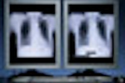
"Chest radiographs of all patients were obtained using a flat-panel digital chest system (Revolution XQ/I, GE Healthcare, Chalfont St. Giles, U.K.), based on a CsI scintillator and an amorphous silicon photodiode-transistor array. The detector has an image size of 41 x 41 cm and a pixel dimension of 0.2 x 0.2 mm. The dual-energy examination consisted of standard posteroanterior (PA) radiographs as well as subtracted soft-tissue and bone images" (European Radiology, September 2008, Vol. 18:9, pp. 1831-1839).
"Dual-energy images were acquired applying a dual-exposure technique within 200 msec. The imaging parameters included a 120 kV and 2 mAs image at a speed equivalent to approximately 400 and a 60 kV and 8 mAs image at a speed equivalent to approximately 1,000," Dr. Röhl and colleagues wrote.
Nodule status was determined by either histological evaluation or follow-up CT. The authors considered an increase in nodule diameter of more than 25% on CT at six-month follow-up to indicate malignancy. They used the standard radiography for the high-kilo image so that the only additional image was the low-kilo image, which increased the total dose by 14%.
"The cumulative sensitivity of chest radiography in conjunction with dual energy was 43%, specificity was 55%. For digital radiography alone, sensitivity was 35% and specificity was 83%," Röhl and his team wrote. "For the dual-energy system, positive predictive value was 58%, and negative predictive value was 66% compared to the digital radiography with a positive predictive value of 59% and a negative predictive value of 65%," they wrote.
"CsI-based flat-panel dual-exposure dual-energy imaging added to standard chest radiography did not show statistically significant improvement for the detection of pulmonary nodules, nor the identification of calcifications, nor the determination of malignancy," the authors concluded. "Even though the relative sensitivity of the subtraction technique (39, 9%) proved to be slightly higher in comparison with standard chest radiography (34, 9%), no significant improvement was reached for the overall detection of pulmonary nodules."
"CsI-Detector-Based Dual-Exposure Dual Energy in Chest Radiography for Lung Nodule Detection: Results of an International Multicenter Trial"
R. Röhl et al
Universitätsklinikum Magdeburg, Klinik för Radiologie und Nuklearmedizin, Leipzigerstr. 44, 39120 Magdeburg, Germany
European Radiology 2008; 18:1831-1839
By Radiology Review
December 26, 2008
Copyright © 2008 AuntMinnie.com