When imaging facilities debate whether to install digital x-ray technology, most are primarily interested in the workflow benefits that derive from bringing radiography into the digital age. But there is also a side benefit to digital x-ray, in terms of advanced applications such as computer-aided detection (CAD) or dual-energy subtraction that either are much more cumbersome or aren't possible at all with conventional analog radiography.
In the case of dual-energy subtraction, it's been available for almost 15 years as a method of differentiating between soft tissue and bone to discover potentially cancerous nodules. The proliferation of computed radiography (CR) and digital radiography (DR) is making dual-energy subtraction more readily accessible, and more clinicians and radiologists are making the technique part of their routines. But other healthcare providers wonder if dual-energy subtraction is worth the extra time it takes to produce and read the studies.
Dr. Anthony Garritano, a visiting radiologist at the Department of Veterans Affairs Medical Center in Bay Pines, FL, uses six images as part of his dual-energy subtraction process with CR. After the two initial images -- a standard posteroanterior (PA) and lateral (frontal and side) view -- are obtained, Garritano uses CR to create four additional images -- a PA and lateral view of the tissue, and an additional PA and lateral of bone structures -- which adds no time or expense to the process. The value of the varying perspectives is detecting abnormalities, lesions, or cancers that could be hidden behind a bone.
"When you are looking at x-rays, you are looking through everything -- the bone, the lungs -- on chest x-rays," Garritano said. "It is quite possible for a tiny lesion to be behind a bone, but not dense enough to see on a routine x-ray until you get the bone out of the way."
When dual-energy subtraction eliminates the bone structure, small cancers can be seen. "If it weren't for dual-energy subtraction imaging, we would not have seen those (lesions) until they got bigger, and then you're running into problems with the patient, because the cancer has been growing," Garritano said.
In other cases, there may be a small lesion between the ribs. If the lesion is smaller than 5 mm, it may go undetected on a standard x-ray image. Another dual-energy subtraction benefit is in detecting suspicious rib fractures or rib lesions that are quite small, possibly invisible to standard rib x-rays, because of the obstruction of other soft tissue.
When looking at the possibility of a bone tumor in an upper or lower extremity, such as the thigh bone or tibia, dual-energy subtraction also aids in detecting whether the tumor is in the bone itself or the soft tissue around the bone.
Dual-energy subtraction may be most beneficial in determining if a lesion or nodule contains calcium. "If there is calcium in it, chances are it is not malignant," Garritano said. "With dual-energy, if you remove the soft tissue, you see a dense, dark area that represents calcium. If you subtract the bone structure and the calcium deposit goes away and all you see is the lesion, then you know it has no calcium."
Cardiac applications
Dr. Robert Gilkeson, the director of cardiothoracic imaging at University Hospitals in Cleveland, has been using dual-energy subtraction with DR for the last four years. Currently, it is a part of the hospital's routine for any kind of chest x-ray.
Gilkeson sees dual-energy subtraction as a potentially useful tool in detecting cardiovascular disease, primarily in the viewing of calcium in coronary arteries and serving as a "screening tool or opportunity to look at coronary artery disease in a noninvasive way."
 |
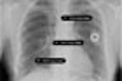
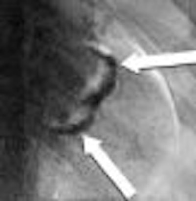
| Above: PA chest radiograph of an 86-year-old female. Below: Dual-energy subtraction image of figure above. The subtle left upper lobe mass is demonstrated (arrow). |
 |
"There is no doubt that you see calcium in coronary arteries in dual-energy that you don't see off a standard x-ray," he said.
At the very least, Gilkeson added, the detection of calcium should prompt a radiologist to alert a referring physician to the possibility of coronary artery disease in a patient with chest pain.
 |
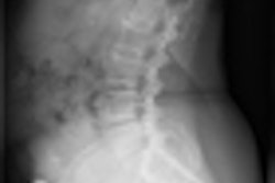
| Dual-energy bone image of figures above. The mitral annular calcifications are demonstrated (arrows). All images courtesy of Dr. Robert Gilkeson and medical student Sonali Mehandru, University Hospitals in Cleveland. |
Garritano, however, is not as convinced of dual-energy subtraction in coronary imaging. "If you are looking for hardening of coronary arteries with calcium, the calcium is so small that it may be enough to block an artery, but not (large) enough to be seen on x-ray," he said. "The only way calcified coronary arteries can be visualized is with CT."
Possible limitations
Gilkeson co-authored a 2006 paper on dual-energy subtraction in the Journal of Thoracic Imaging (November 2006, Vol. 21, No. 4, pp. 303-313), in which he and Dr. Peter B. Sachs of Harvard Medical School in Boston discussed potential limitations with dual-energy digital chest radiography. While the research had positive reviews for dual-energy subtraction, there were some caveats.
The study noted how a standard chest x-ray is performed, followed by a lower-energy image of 60 kV taken 150 msec later. With the acquisition of a second lower-energy image, the paper reported that there could be a "somewhat noisier display in areas of increased soft tissue, such as the pleura/chest wall and lung base/diaphragm interface."
In addition, despite the short amount of time between image acquisition (150 msec), if a patient has difficulty holding his or her breath or if there is any change in position of the chest, there are "significant artifacts" with dual-energy subtraction, according to Gilkeson. "The standard x-ray looks fine, but, if there is a lot of breathing," it will adversely affect image results, he said.
Will it catch on?
So why isn't dual-energy subtraction used more frequently? Gilkeson believes that one reason is that busy radiologists may not want to add to their already voluminous workload. "Like anything, there is a learning curve; you have to look at a few more images," he said.
From a workflow perspective, Gilkeson estimated the additional images from dual-energy subtraction may add another five to 10 seconds to an x-ray reading.
In addition, some areas are difficult to see because of the soft tissue. Areas behind the heart and around the diaphragm can be problematic, but, in time, the obstacles are overcome. "Once you recognize the artifacts, it is easy to discard them as artifacts," Gilkeson noted.
Both Garritano and Gilkeson discount the issue of radiation exposure due to the additional image acquisition with dual-energy subtraction. The majority of the radiation dose is expended in the lateral film, and the second PA exposure has less radiation. Compared to radiation from a standard film-screen chest x-ray, the dual-energy exam may result in approximately 5% more radiation. "Given the benefits of the (dual-energy) technology, I think that 5% amount of radiation is very well worth it," Gilkeson said.
Garritano leans toward dual-energy subtraction as the first step to cancer detection, in part, because the acquisition of two images offers less radiation than multislice CT. "If you did see a small lesion, then the next step would be a CT (scan), because you are looking to see if the lesion has spread," Garritano said. "Where it (CT) helps in the dual-energy subtraction technique is saving cases where a tiny lesion would be missed and the cancer would grow."
By Wayne Forrest
AuntMinnie.com staff writer
February 22, 2007
Related Reading
Chest radiographs negative for one in five acute heart failure patients, January 13, 2006
I-ELCAP data suggest need to work up secondary lung lesions, January 9, 2006
Chest x-ray after thoracentesis adds little new information, December 26, 2005
Lung PET rules when CT is equivocal, discordant, July 12, 2005
Copyright © 2007 AuntMinnie.com