In a study of more than 250 computed radiography (CR) systems from three vendors, a group of researchers from Vancouver General Hospital in British Columbia have found that premature wear of cassettes and image plates can cause image artifacts.
The report, published in the Journal of Digital Imaging by the Society for Imaging Informatics in Medicine (SIIM, formerly SCAR), was prompted by a rise in complaints of image artifacts in patient images 12 to 18 months after installation of new CR systems (JDI, September 2006, Vol. 19:3, pp. 226-239).
The team, led by radiologic technologist Kevin Hammerstrom, evaluated 197 CR cassettes and imaging plates from Fujifilm Medical Systems USA of Stamford, CT; 35 from Agfa HealthCare of Greenville, SC; and 37 from Eastman Kodak Health Group of Rochester, NY.
Of the cassettes and image plates reviewed in the study, the maximum number of times any cassette and image plate had been read was approximately 10,000 for an Agfa system, 7,500 for Fuji, and 3,600 for Kodak. The average number of times that Fuji cassettes and image plates were read was approximately 1,560. Physical damage to the exterior of the Agfa and Fuji cassettes was more extensive on those used more frequently.
Overall, the researchers found that cassettes in the sizes of 18 x 24 cm, 24 x 30 cm, and 35 x 35 cm were in good condition, compared to 35 x 43-cm cassettes, which were used more often.
Upon further review
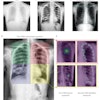
The CR inspections discovered various issues in each vendor's products, from the accumulation of dust in grooves around the edges of some devices to a fractured cassette and minor damage to some cassette hinges. The study also noted what the authors described as "black specks" on the phosphor surface of imaging plates from all vendors, with many of the specks visible on the radiographic flat-field images.
One vendor's imaging plate appeared to have a "fine-grained, dark-colored dust" on the phosphor surface that caused the flat-field radiographic image to appear lighter than an area from where the dust was removed. The authors concluded that the dust most likely resulted from repeated friction between the aluminum surfaces of the imaging plate's backing and the open edge of the cassette whenever the imaging plate was removed and reinserted during the reading process.
Random scratches also were found on the phosphor surface of all three vendors' imaging plates. The researchers surmised that the scratches were most likely caused by the phosphor surface coming into contact with interior components of the CR system during manual removal of the imaging plate after jams had occurred, or were caused by contact between the phosphor surface and the open end of the cassette when the imaging plate was transported into and out of the cassette.
Other notable findings included the yellowing of the phosphor edges on 39.5% or more of each size of Fuji image plate, on approximately 30% of 35 x 35-cm and 35 x 43-cm Agfa imaging plates, and on approximately 33% of 35 x 43-cm Kodak imaging plates.
Maintenance recommendations
The authors concluded that the CR devices' cassettes and imaging plates "are the greatest source of image artifacts," and that many image artifacts "were the direct result of premature wear of the cassettes and imaging plates, some caused by flaws in their design." They recommended a quality control and regular maintenance program of cleaning the cassettes and imaging plates, as well as visual and radiographic image inspection. Even the simple removal of dust or dirt particulates can prevent the deterioration of image quality, they stated.
Also, the group advised radiology departments to document the physical condition and imaging performance of each cassette and imaging plate to monitor the progress and location of deterioration to help prevent it in the future.
By Wayne Forrest
AuntMinnie.com staff writer
September 21, 2006
Related Reading
Standardized testing enables CR and DR cohabitation, August 11, 2005
Ortho group stumbles with DR, completes conversion to digital CR, May 27, 2005
New research shows CR more cost-effective than DR, February 28, 2005
Study compares eight digital x-ray systems in clinical setting, December 24, 2004
Computed radiography's (not so surprising) resilience, September 16, 2004
Copyright © 2006 AuntMinnie.com