Loss of blood supply, infections, and mechanical instability are some of the factors that can lead to nonunion of damaged bones. Inadequate bone healing can adversely affect patient management. Researchers from Austria found that multidetector-row CT (MDCT), and not x-ray, was the most efficient way to monitor bone healing.
Generally, two-view radiographs have been used in this setting, but they are not a dependable test, wrote Dr. Christian Krestan and colleagues in the American Journal of Roentgenology. Standard CT has also been used to evaluate bone production, but the results can be influenced by scanner type and imaging parameters (AJR, June 2006, Vol. 186:6, pp. 1754-1760).
Kresten's group, from the departments of radiology and orthopedic surgery at the Medical University of Vienna, conducted a retrospective study on 43 patients with a history of fractures, spondylodesis, and other orthopedic conditions. MDCT was performed on a 16-slice scanner (Mx8000 IDT, Philips Medical Systems, Andover, MA) with multiplanar reconstructions (MPRs).
Digital x-rays (Multix FD, Siemens Medical Solutions, Malvern, PA) were done in two views: the lumbar spine and hip at 70 kVp and 50 mAs, and the ankle joint at 555 kVp and 6 mAs. The system featured a flat-panel detector with a cesium iodide amorphous layer (Trixell Pixium 4600, Siemens Medical Solutions).
 |
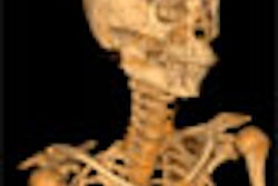
| A 65-year-old woman with history of L4-L5 fusion and anterolisthesis of L3-L7. Sagittal multiplanar reconstruction shows complete fusion (arrow). |
Digital radiographs and MDCT images were analyzed independently of each other by two musculoskeletal radiologists. According to the results, MDCT showed no evidence of bone bridging in 32.6% of the cases. Partial fusion could be seen on MDCT in 53.5% of the cases and full fusion in 14%. More than half the patients had metal implants, which lead to minor artifacts on MDCT, the group stated.
Overall agreement between the two modalities was found in 63% of the patients. Digital x-ray overestimated the bone healing process in 19% of the patients and underestimated it in 19% as well.
 |
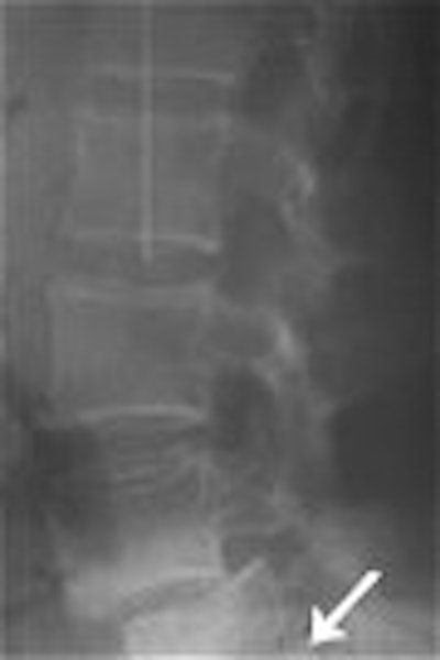
| Lateral radiograph shows partial fusion. Krestan CR, Helge H, Vasilevska V, Weber M, Schueller G, Imhof H , and Czerny C, "MDCT Versus Digital Radiography in the Evaluation of Bone Healing in Orthopedic Patients" (AJR 2006; 186:1754-1760). |
In 16 patients, MDCT altered the diagnosis initially made on x-ray and changed the initial diagnosis with a good diagnostic confidence level at a higher percentage (37.5%-50%), the authors stated. In 26.5% of the cases, the diagnostic confidence was rated as reliable.
The authors concluded that digital x-ray was an unreliable diagnostic technique and could not serve as a sufficient method to exclude delayed healing and nonunion. Ultimately, the orthopedic surgeons relied on the final MDCT results before proceeding with treatment, they added.
By Shalmali Pal
AuntMinnie.com staff writer
May 31, 2006
Related Reading
Bone marrow edema on preop MR correlates with vertebroplasty success, May 18, 2006
Course of stress fractures in the juvenile skeletal system often prolonged, April 25, 2006
CT more sensitive than spinal fracture x-ray, but has limits, January 30, 2006
Copyright © 2006 AuntMinnie.com