Ever since it appeared on the clinical scene, functional-based breast-specific gamma imaging (BSGI) has sparked the question of whether it is sensitive enough to replace other adjunctive modalities used with mammography for high-risk patients, such as MRI or ultrasound.
In June, Dr. Rachel Brem and colleagues at George Washington University in Washington, DC, published work in Radiology that showed BSGI to be as sensitive as MRI for detecting most cancers, and more sensitive for detecting ductal carcinoma in situ (DCIS). And in a presentation at the 2008 RSNA meeting in Chicago, she offered data that indicate BSGI can reveal cancers that mammography misses.
Now, in a new study, Brem and colleagues compared BSGI's sensitivity in detecting invasive lobular carcinomas to that of mammography, sonography, and MRI. The team's findings were published in this month's issue of the American Journal of Roentgenology (February 2009, Vol. 192:2, pp. 379-383).
The study included 26 women between the ages of 46 and 82 who had biopsy-proven invasive lobular cancer. All had undergone mammography and BSGI, and the results of sonography and MRI, if performed, and the pathologic tumor size were also included.
Twenty-eight biopsy-proven invasive lobular carcinomas were found in the 26 women. The mean pathologic size of the tumors was 22.3 mm, and mammographic findings were negative for six cancers. Abnormal mammographic findings, found in 22 of the 28 cancers, included:
- 13 (59%) asymmetric densities
- 4 (18%) architectural distortions
- 5 (23%) spiculated masses
For seven of the 22 patients (32%), the invasive lobular carcinoma was revealed as microcalcifications. Overall, mammography had a sensitivity rate of 79% for detecting these cancers.
Twenty-five women also had sonography, and Brem and colleagues found 17 focal hypoechoic areas. Eight patients had negative sonography exams; of these eight lesions not detected by sonography, BSGI missed one. The sensitivity of sonography for detecting these cancers was 68%.
Of the 26 women included in the study, 12 also had MRI exams. Ten of 12 lesions, or 83.3%, enhanced after the injection of gadolinium contrast, four of which had not been caught by mammography. The mean size of the cancers detected by MRI was 19.9 mm, and the sensitivity of MRI for finding invasive lobular carcinoma was 83%.
Finally, BSGI showed an increase in radiotracer uptake in 26 of 28 cancers, for a sensitivity of 93%. The mean size of lesions detected by BSGI was 20.3 mm; the smallest invasive lobular carcinoma BSGI found was 2 mm. The two lesions BSGI did not find were 5 mm and 90 mm. The technology found six cancers that mammography did not, and two cancers for which MRI findings were negative (5 mm and 40 mm).
Test results for the detection of invasive lobular carcinomas
using each imaging technique
|
|||||||||||||||||||||||||||
| Table courtesy of the American Roentgen Ray Society. | |||||||||||||||||||||||||||
"There was a nonsignificant trend for BSGI to have a higher sensitivity for the detection of invasive lobular carcinoma than mammography, sonography, or MRI," the authors wrote. Additional and larger studies are needed to further investigate this trend, they noted.
 |
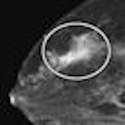
| Breast images of 46-year-old woman with biopsy-proven bilateral lobular carcinoma who presented with palpable left breast mass. MRI and breast-specific gamma imaging findings were positive. Mammogram of right breast (not shown) was negative, whereas mammogram of left breast (not shown) showed pleomorphic calcifications in upper outer quadrant. Above and below, contrast-enhanced MR images of right breast show spiculated mass (circle) at 12-o'clock position. All images courtesy of the American Roentgen Ray Society. |
 |
 |
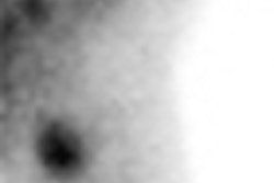
| Craniocaudal breast-specific gamma images of left (above left) and right (above right) breasts and mediolateral oblique images of left (below left) and right (below right) breasts show increased uptake at 12-o'clock position in right breast (arrows) as well as increased uptake in upper outer quadrant of left breast. |
 |
In addition to its greater specificity over MRI, BSGI has other advantages, according to the authors:
- The exam is performed with the patient sitting comfortably rather than being confined in an MRI scanner.
- The BSGI exam generates from four to 16 images, at most, compared with hundreds or even thousands of images for a breast MRI exam.
- The cost of a BSGI examination tends to be less than that of breast MRI.
- With the increasing concern of renal complications with the administration of gadolinium, the IV injection of technetium-99m sestamibi has not been reported to be associated with significant complications.
At 93%, BSGI has the highest sensitivity for the detection of invasive lobular cancer as compared to mammography, sonography, and MRI, Brem and colleagues concluded. It's an "effective technique that should be used to evaluate patients with suspected cancer and has a promising role in the diagnosis of invasive lobular carcinoma," they wrote.
By Kate Madden Yee
AuntMinnie.com staff writer
February 11, 2009
Related Reading
Breast-specific gamma imaging reveals unseen cancers, December 4, 2008
Dual-head gamma camera increases breast lesion detection, November 28, 2008
BSGI sensitive in detecting cancer, study says, May 30, 2008
Breast gamma imaging spots DCIS better than mammo, MR, August 13, 2007
ARRS study: Scintimammography shows high sensitivity in breast cancer detection, May 9, 2007
Copyright © 2009 AuntMinnie.com