HOUSTON - Duke University researchers have begun the first patient studies of a hybrid SPECT/CT mammography system, and the unit has several advantages over conventional breast imaging technology, according to their findings.
The system was tested for the first time last month on patients diagnosed through biopsy, according to Martin Tornai, Ph.D., an assistant professor at Duke University's department of radiology in Durham, NC, and inventor of the technology, and Priti Madhav, a biomedical engineer who fused the systems together. They presented their results at the 2008 American Association of Physicists in Medicine (AAPM) meeting.
"The hybrid combines two separate datasets to get a single image with functional and anatomical representation," Madhav said.
The area of biopsy, tracers placed around the tumor, and views into the chest wall were all plainly depicted in a 3D image Madhav presented of a patient with breast implants. The analysis included comparisons of images from four patients imaged with SPECT, two with CT, and one with both. The separate images were fused using markers placed at superior, inferior, lateral, and medial points.
 |
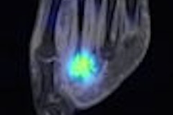
| Reconstructed coronal (left) and sagittal (right) fused SPECT/CT image slices (hot scale = SPECT, black and white scale = CT) from a patient's breast. Suspected cancer shows region of enhanced sestamibi uptake (indicated with green hashmarks). Cancer was confirmed with biopsy and MRI as being ductal carcinoma in situ. A biopsy clip is seen in the upper region of the sagittal slice (arrow), just above the sestamibi uptake in the lesion. SPECT bright areas outside the breast are external fiducial markers and areas above the breast are due to the myocardium. All images courtesy of Priti Madhav. |
Tornai told AuntMinnie.com that the hybrid imaging system is an especially good solution for patients with breast implants because it does not rely on compression, which can cause implant eruption.
The researchers said the first results in human subjects offer preliminarily confirmation that the system's 3D capability yields better images than a screening mammogram at 25% lower dose. "The dose is two-thirds that of a screening mammogram," Tornai said. "The breast is very radiosensitive, and this lessens exposure to unnecessary radiation."
Comfort also factored into the design. Madhav customized a bed for positioning patients in a prone position, with the breasts hanging pendant. The device acquires 3D data underneath the bed in an open field-of-view. The prototype bed is lined with neoprene and redlined to reduce scatter.
The SPECT component of the system acquires the 3D image through complex trajectories around the breast, while the CT subsystem, a monochromatic, cone-beam x-ray source, makes one circular pass, thereby limiting radiation dose and increasing contrast.
 |
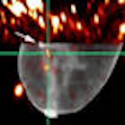
| Dedicated hybrid SPECT/CT imaging system with a customized patient bed. The SPECT camera is placed orthogonal to the CT tube and CT detector. An uncompressed breast phantom is placed in the common field-of-view of the two systems. The SPECT system can move fully in 3D motion while the CT system is restricted to a circular rotation. |
|
Video courtesy of Duke University Medical Center Multi-Modality Imaging Lab. Having trouble viewing this clip? Click here to view full-size clip or to change format. |
One disadvantage of the system over conventional mammography is the exam time, which is longer for both the SPECT and CT portions. Madhav said the 10-minute SPECT acquisition time can't be shortened, but she expects to reduce the duration of the CT portion from 4.5 minutes to 1.8 minutes.
If a clinical product is developed, the system may -- or may not -- become the first dedicated SPECT/CT system for breast imaging. Tornai said two other research teams in the U.S. are also working on combined SPECT/CT technology.
Duke University and a university spin-off lab Tornai started with funding from the National Institutes of Health (NIH) have jointly applied for a technology patent for the system.
By Kathlyn Stone
AuntMinnie.com contributing writer
July 30, 2008
Related Reading
Studies support breast CT's ability to heighten tissue contrast, resolution, January 25, 2008
Duke MMIL designs novel SPECT/CT system for 3D breast imaging, July 18, 2007
Imaging start-up to install conebeam CT breast imaging prototype, May 1, 2006
Breast CT developers aim to 'revolutionize' mammography, December 20, 2005
Researchers make inroads with breast CT, gear up for clinical test, July 25, 2005
Copyright © 2008 AuntMinnie.com