BOSTON - The teniae coli, three thick bundles of muscle running along the outer longitudinal layer of the colon's muscularis externa, have a big job to do. Together they enable colon segments to contract independently, one after the other, squeezing contents along the lumen toward the distal end.
As if that weren't enough, researchers at the National Institutes of Health (NIH) in Bethesda, MD, have given the teniae new job responsibilities -- as guides for virtual colonoscopy readers navigating the inside of the colon.
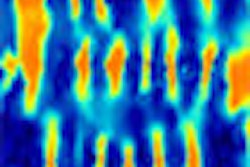
On CT colonography datasets the teniae coli can be identified as bands of smooth muscle extending parallel to the axis of the colon. Once detected, colorized and digitized, they present a rather elegant anatomic solution to the perennially difficult job of registering polyp locations. In future CAD systems, the teniae coli could even help localize and register polyps found on the corresponding prone and supine scans of VC studies.
"The motivation behind this project was to determine if we could use the teniae coli ... to aid in polyp localization and navigation," said Dr. Ron Summers, Ph.D., at the 2005 International Symposium on Virtual Colonoscopy. The teniae coli "are instrumental in peristalsis and propulsion of fecal matter," he said. "They are named by their position on the transverse colon and by the structures that attach to them."
Individually the muscles are known as tenia omentalis (TO), attached to the omentum; the tenia mesocolica (TM), attached to the mesocolon; and the tenia libera (TL), or "free" tenia, so named because it has no mesenteric attachment.
NIH investigators led by Dave Roy, with colleagues Adam Huang, Ph.D.; Marek Franaszek, Ph.D.; and Summers, developed a three-step semiautomated method to detect the teniae coli following the initial reconstruction of CT colonography (VC) datasets.
"Step one is to choose steps along the TO and construct a path -- this method is currently a manual path in this early stage of the algorithm," said Summers, who is a senior investigator and staff radiologist at the NIH.
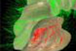
Step two is to construct a path through those points, he said. Step three uses the path along the TO to automatically derive paths along the remaining two teniae coli. Step three is achieved by virtually cutting open the colon surface along the length of the TO and flattening it out into a rectangular sheet. Then two lines are drawn longitudinally to divide the rectangle into thirds. Finally, the lines are mapped back into the original CT dataset, thereby approximating the position of the remaining two teniae: the TM and the TL.
What's left is a kind of color-coded highway down the colonic lumen, where the TO is always located at high noon, the TL at 8 o'clock, and the TN at 4 o'clock, no matter how the colon twists and turns in the course of an endoscopic fly-though. The effect is quite steadying.
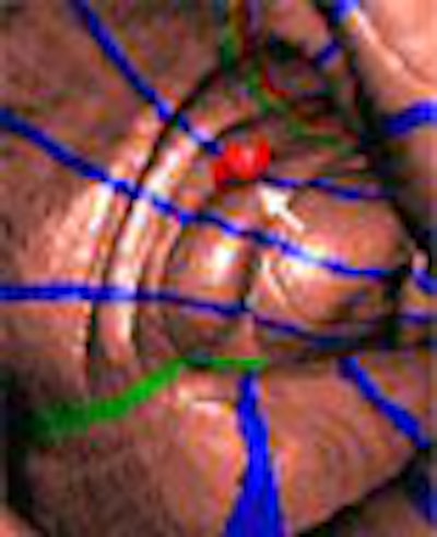
"This is the TL, which is this yellow line ... and you'll notice that although these are approximations to the course of the teniae -- for example the TL is actually slightly above where this line is drawn -- it's a good approximation," Summers said. "Haustral folds are indicated in blue."
How to put the markings to good use? To this end the team examined five patients with colonoscopy-proven polyps to evaluate the algorithm's ability to derive the teniae coli paths in each dataset, and assess its ability to create a polyp localization system, Summers said.
After the TO path was identified manually in each dataset, the automated algorithm chose the paths of the other two teniae, verified by the investigators' visual inspection. Next the polyps were identified as being in one of six possible circumferential positions with respect to the teniae coli.
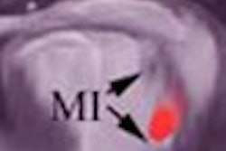
"We looked at five patients having polyps on both the prone and supine and scans, and we recorded and compared the circumferential locations of these polyps," Summers said. "Here's a polyp near the tenia mesocolica on the supine scan, and on the prone scan you can see it's quite close to the TM on both views."
The results shows that polyp positions were relatively fixed with respect to the teniae, and all were localized in the same circumferential position on the supine and prone datasets, Summers said.
 |
| Synchronous view of a colonic polyp in the supine and prone positions. The polyp in the picture is a 7-mm round adenoma located at the hepatic flexure. The left panel is the supine view; at right is the prone view. Tenia omentalis is the red-yellow-green stripe; tenia mesocolica is colored in green; tenia libera is yellow. Images courtesy of Adam Huang, Ph.D., and Dr. Ron Summers. |
In endoluminal navigation of a dataset, "the idea is that the TO is a guidewire, and by keeping the TO at the 12 o'clock position for the supine and prone scans, and registering the colons also along their length, you have both longitudinal and circumferential registration of the colon, which is our ultimate goal for registration," he said. "We're finding polyps we couldn't see before."
The circumferential positioning system was successful in all 10 datasets (five patients supine and prone) the researchers wrote in their abstract. In future CAD systems, "the tenia omentalis can serve as a guidewire for oriented and registered VC fly-throughs," Summers said.
By Eric Barnes
AuntMinnie.com staff writer
October 19, 2005
Related Reading
VC safe, surveys show, but not without incident, October 18, 2005
Colonoscopy often inaccurate in localizing colorectal cancer, October 18, 2005
New VC reading schemes could solve old problems, October 13, 2005
'Filet view' VC software slices reading time, October 6, 2005
VC path-planning technique maximizes observable mucosa, August 30, 2005
Copyright © 2005 AuntMinnie.com