BOSTON - U.S. military researchers were all they could be at this week's International Symposium on Virtual Colonoscopy. At Monday's opening sessions, Navy radiologist Dr. Perry Pickhardt reported the most accurate results ever seen in a large virtual colonoscopy trial.
The study raised eyebrows a bit higher with Pickhardt's assertion that primary 3D interpretation with 2D correlation was key to the group's success. Most virtual colonoscopy providers have taken the opposite approach: 2-D slice review followed by 3D for problem-solving.
The prospective trial of 1,233 asymptomatic patients was conducted between May 2002 and June 2003 at the National Naval Medical Center in Bethesda, MD; the Walter Reed Army Medical Center in Washington, DC; and the Naval Medical Center in San Diego.
Pickhardt, along with Dr. J. Richard Choi, Dr. Inku Hwang, and colleagues at the three centers, had also raised the stakes by excluding the high-risk patients that have formed the core of most virtual colonoscopy trials, relying instead on a screening population of mostly average-risk adults (728 men, 505 women ages 40-79, mean age 57.8 years) whose low prevalence of colorectal polyps and cancers could presumably make lesions harder to detect. Only 32 patients had a family history of colorectal cancer, constituting a tiny subset of patients with above-average risk. This subset included the only patients under age 50.
Patients with rectal bleeding, a history of prior polyps or cancer, or any history of colorectal cancer screening tests in the previous 10 years were excluded, Pickhardt said.
Virtual colonoscopy was performed by six radiologists who had undergone virtual colonoscopy training and performed between 25 and 150 studies each. Seventeen experienced endoscopists performed the same-day conventional colonoscopy on all 1,233 patients.
Prior to CT scanning, all subjects underwent a 24-hour purgative bowel cleansing with two 45-ml doses of phosphosoda and 10 mg of bisacodyl, along with two 250-ml doses of 2.1% barium contrast material to mark any fecal residue. Patients self-insufflated the colon with room air to tolerance, which was pretty high in some of the ex-Marines, Pickhardt remarked later, though a few patients were gun-shy.
CT images were obtained on either a four- or eight-slice multidetector-row scanner using 2.5-mm or 1.25-mm collimation, respectively. Both prone and supine scans were performed using 15 mm/sec table speed, 1-mm reconstruction intervals, 100 mAs and 120 kVp.
The group used a V3D-Colon workstation (Viatronix, Stony Brook, NY) for primary 3D interpretation, reserving 2D for problem-solving and correlation of all positive results. In another twist, conventional colonoscopy results were segmentally unblinded, allowing for the assessment of any false negatives in conventional colonoscopy, which served as the reference standard. In a rigorous threshold-based analysis, the results were matched both by polyp and by patient.
Surprising results
At conventional colonoscopy the group detected a total of 1,310 polyps in 1,233 patients, including 344 (26.2%) 6 mm or larger, 210 of which (61%) were adenomatous. Only two of 544 total adenomatous polyps were malignant, Pickhardt said.
In by-polyp matching with conventional colonoscopy, the most rigorous measure of accuracy, VC showed sensitivity of 91.5% (54/59) for polyps 6 mm and larger, handily surpassing the 88.1% (52/59) demonstrated by conventional colonoscopy. Moreover, virtual colonoscopy detected both adenomas, while only one was seen on conventional colonoscopy until the VC results were unblinded and the second adenoma was located retrospectively.
 |
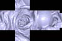
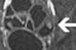
| Eight-millimeter adenomatous polyp in ascending colon seen in 3D endoluminal view (above); in translucent view used to distinguish polyp from stool (below); and in corresponding 2D axial view (bottom). This lesion was missed at prospective conventional colonoscopy, but found after unblinding of VC results. Images courtesy of Dr. Perry Pickhardt. |
 |
 |
Using a threshold-based analysis, VC's sensitivity was 88.7% for polyps 6 mm and larger, 93.9% for polyps 8 mm and larger, and 93.8% for lesions 10 mm and larger. Conventional colonoscopy's sensitivity trended slightly lower: 92.3%, 91.5% and 87.5%, respectively.
VC's accuracy kept improving as the polyp threshold size rose: 80.9% for polyps 6 mm and larger, 92.3% for polyps 8 mm and larger, and 95.9% for polyps 10 mm and larger. As for specificity, virtual colonoscopy was 79.6% specific for polyps 6 mm and larger, 92.2% specific for polyps 8 mm and larger, and 96% specific for polyps 10 mm and larger, Pickhardt said.
"So what are the implications of our study? I believe that use of the primary 3D approach should establish virtual colonoscopy as a front-line screening option," Pickhardt said. It compares favorably with the reference standard, he said, and performs optimally for polyps 8 mm and larger.
"One of the reasons for ... our results with the primary 3D approach ... is certainly easier polyp conspicuity and better fold depiction.... If you start with an endoluminal fly-through, you're just going to find a lot more polyps than with axial images," he said.
In addition, the use of segmental unblinding of VC results turned what would have been false-positive VC results into false-negative conventional colonoscopy results.
When the colonoscope was being withdrawn in conventional colonoscopy, it had a tendency to slip past a few polyps, especially those located on or near haustral folds, he said later. But these polyps tended not to be missed in virtual colonoscopy, and thanks to segmental unblinding of VC results, could be picked up retrospectively with the colonoscope. The use of multidetector-row scanners with thinner collimation also improved VC's performance, he said.
Indeed, the group's demonstrated sensitivities in the low 90s were more than 10 points higher than the best results in previous studies; the tricky part was explaining how it had occurred so consistently and in such a large patient population. The radiologists also showed high interobserver agreement, with fairly consistent mean interpretation times ranging from 15.9 to 24 minutes.
In a later discussion, some attendees challenged the idea that 3D primary interpretation was the main driver of the outstanding results, noting that previous studies had not demonstrated a clear advantage to 3D evaluation.
Some suggested that a combination of positive factors, in addition to the Viatronix software, may have primed the pump for success. Factors such as the barium fecal tagging, the thin-section imaging, and excellent colonic distension throughout the cohort may have been just as important as primary 3D interpretation, they said. Perhaps the readers were simply an outstanding group of radiologists. And could it be that the patients, on the young side for virtual colonoscopy subjects, were motivated to do a better-than-average job with colon cleansing and self-insufflation?
Maybe 3D reading makes the entire learning curve easier to climb, as one attendee suggested. But in that case, could over-reliance on 3D be a potential barrier to the development of essential 2D reading skills?
Pickhardt allowed that any of these factors might have helped. But he said the yearlong project had convinced the group that the workstation, designed for primary 3D interpretation, had played the biggest role in facilitating reading.
By Eric BarnesAuntMinnie.com staff writer
October 15, 2003
Related Reading
Virtual colonoscopy not yet an adequate screening tool for colorectal cancer, October 13, 2003
Group favors 3-D primary read in major virtual colonoscopy trial, October 2, 2003
Virtual colonoscopy: 2-D vs. 3-D primary read, June 3, 2002
Copyright © 2003 AuntMinnie.com