Prostate cancer is the most frequently occurring cancer in men. For individuals categorized as high-risk patients, detection or exclusion of osseous metastatic disease is of high clinical importance in the management of their prostate cancer.
"Newly diagnosed patients with localized disease and no metastases may benefit from radical localized curative treatment, in contrast with patients who bear metastases, in whom early initiation of androgen withdrawal and bisphosphonate therapy and withholding of unnecessary radical therapy such as radiotherapy is the appropriate treatment approach," wrote researchers from Israel in a recent issue of the Journal of Nuclear Medicine (February 2006, Vol. 47:2, pp. 287-297).
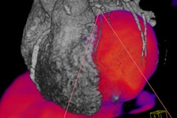
The scientists, from Tel Aviv and Kfar Saba, Israel, conducted a prospective study comparing the detection of bone metastases by technetium-99m methylene diphosphonate (Tc-99m MDP) planar bone scintigraphy, single and multiple field-of-view (FOV) SPECT, F-18 fluoride PET, and F-18 fluoride PET/CT in patients with high-risk prostate cancer.
The researchers selected the radiotracer F-18 fluoride because it is a bone-seeking positron-emitting agent that is characterized by a twofold higher bone uptake than Tc-99m MDP. However, it is not tumor-specific and is prone to delivering a high false-negative rate, which can be mitigated by lesion correlation with CT or MR imaging, they noted.
The team conducted bone scintigraphy, SPECT, and PET/CT exams on the same day on patients who had been diagnosed with prostate cancer and had a high risk for bone metastases. The group examined 44 patients with a mean age of 71.6 years; 25 of the patients were newly diagnosed, while 19 were referred for evaluation of suspected recurrence or progression.
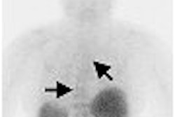
Planar bone scintigraphy of the axial skeleton was the first exam performed, and images were acquired two to three hours after injection of 25 mCi of Tc-99m MDP using a Discovery (GE Healthcare Chalfont St. Giles, U.K.) dual-head gamma camera. The researchers performed single FOV SPECT of the lower thoracic and lumbar spine region on 20 of the men. They also conducted three or four FOV SPECT of the entire axial skeleton on the remaining 24 patients.
The SPECT data was reconstructed using a collimator-detector response method (Evolution software, GE Healthcare). The application incorporates a quantitative model of collimator-detector response function into an iterative reconstruction algorithm, they reported. The use of the algorithm allowed the team to acquire a good-quality SPECT view within eight minutes of acquisition and a SPECT view of the axial skeleton with 24-32 minutes, they wrote.
The PET/CT studies were performed in the afternoon of the same day as the bone scintigraphy and SPECT exams, 60-90 minutes after intravenous administration of 8-12 mCi of F-18 fluoride using a using a Discovery LS (GE Healthcare) PET/CT system.
"Low-dose CT acquisition was performed first with 140 kVp, 80 mAs, 0.8 seconds per CT rotation, a pitch of 6, and a table speed of 22.5 mm per second, without any specific breath-holding instructions," the researchers wrote. "A PET emission scan was performed immediately after acquisition of the CT, without changing the patient's positioning. From five to nine bed positions were performed with an acquisition time of three minutes for each, imaging the skeleton from skull to midthigh."
The PET images were reconstructed and the CT data were used for attenuation correction. All images from the planar bone scintigraphy, SPECT, PET, and PET/CT studies were interpreted by consensus and read separately by nuclear medicine physicians and a radiologist who were blinded to the results, the researchers noted. They found that in 23 of the patients (52%) the prostate cancer had spread to their skeleton, while 21 did not have osseous metastatic disease.
The scientists reported that planar bone scintigraphy had a sensitivity of 35%, specificity of 95%, positive predictive value of 89%, and negative predictive value of 44%. The planar scan and combined single and multiple FOV SPECT had a sensitivity of 39%, specificity of 86%, positive predictive value of 75%, and negative predictive value of 31%.
The multiple FOV SPECT of the entire axial skeleton, when evaluated against its own patient cohort, had a sensitivity of 46%, specificity of 100%, positive predictive value of 100%, and negative predictive value of 61%. F-18 fluoride PET had a sensitivity of 48%, specificity of 95%, positive predictive value of 92%, and negative predictive value of 63%.
The addition of integrated F-18 fluoride PET/CT showed, by far, the strongest potential for detecting skeletal metastatic disease in the high-risk prostate cancer patients with a sensitivity of 57%, specificity of 100%, positive predictive value of 100%, and negative predictive value of 87%.
The authors observed that although F-18 fluoride PET/CT had the highest sensitivity and specificity in their study, it is the least available and most expensive of the techniques that they assessed. As such, they did not recommend that it be introduced as a routine imaging approach of metastatic bone survey in high-risk patients.
Rather, they wrote that "it might be valuable to exploit the high sensitivity and specificity of F-18 fluoride PET/CT in selected cases in which the presence of bone metastasis cannot be definitely confirmed or equally excluded by other imaging modalities."
By Jonathan S. Batchelor
AuntMinnie.com staff writer
March 10, 2006
Related Reading
Alendronate prevents bone loss in men on androgen deprivation therapy, February 28, 2006
Bone SPECT identifies patients who would benefit from facet joint injection, February 15, 2006
SPECT/CT helps clarify ambiguous bone foci, February 9, 2006
Hormone therapy for prostate cancer increases fracture risk, January 6, 2006
SPECT/CT pilots prostate brachytherapy in community setting, August 15, 2005
Copyright © 2006 AuntMinnie.com