There are telltale differences in the chest x-rays of children with H1N1 influenza based on whether patients have a mild or more severe form of the disease, according to an analysis published online December 23 in Radiology.
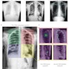
Initial chest x-rays of children with mild cases of H1N1 influenza are often normal but may demonstrate prominent peribronchial markings with hyperinflation. Pediatric patients with a more severe clinical course of H1N1 infection requiring hospitalization have bilateral, symmetric, and multifocal areas of consolidation often associated with ground-glass opacities, according to researchers from Children's Hospital Boston and Harvard Medical School in Boston.
The research team conducted a retrospective study of 108 H1N1 patients who had a chest x-ray performed during a six-month period commencing April 25, 2009. This study group was categorized as follows:
- Children who were discharged after an outpatient clinic or emergency department visit
- Children who were admitted as inpatients for a brief hospitalization
- Children who were admitted as inpatients into intensive care units
Two pediatric radiologists blinded to patient group and lung parenchymal, airway, pleural, hilar, and mediastinal abnormalities systematically reviewed initial chest x-rays. The radiographs were assessed for the presence of pleural effusions or lymphadenopathy, and lung parenchyma and airways were evaluated for patterns, according to lead author Dr. Edward Y. Lee, a pediatric radiologist.
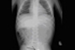
Two-thirds of the children who did not require inpatient hospitalization had normal chest radiographs. Among children who did not require hospitalization but who had abnormal radiographs, the most common finding was prominent peribronchial markings with hyperinflation (17 children).
Because this finding has not been associated with the chest radiographs of adult patients to date, the authors suggested that this may be due to immunologic differences between children and adults. When prominent peribronchial markings and hyperinflation are identified in pediatric chest radiographs, these findings may be associated with an H1N1 influenza infection in children but do not reflect a diagnostic certainty, they added.
The most common findings of chest radiographs of patients requiring hospitalization were bilateral, symmetric, and multifocal areas of consolidation, often associated with ground-glass opacities. This was similar to findings in recent studies of adult patients, the authors reported. However, the radiologists did not observe nodular opacities, reticular opacities, pleural effusion, or lymphadenopathy in any patient.
Related Reading
Chest CT depicts H1N1 better than radiography, October 21, 2009
CT can aid early swine flu diagnosis, October 14, 2009
Virus outbreaks put scrutiny on infection control practices, August 10, 2009
Copyright © 2009 AuntMinnie.com