Scientists at the University of California, San Diego have developed a method to quantify subtle, subregional brain volume loss using MRI, which promises to improve diagnosis and monitoring of Alzheimer's disease, according to the November 16 issue of the Proceedings of the National Academy of Sciences (PNAS).
By applying the techniques to the newly completed dataset of the Alzheimer's Disease Neuroimaging Initiative (ADNI), the scientists demonstrated that such subregional brain volume measurements outperform current measures for tracking the severity of Alzheimer's disease, including cognitive testing and measures of global brain volume loss.
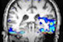
The new methods require at least two brain scans to be performed on the same MRI scanner over a period of several months. Those detected changes in the brain's memory regions, in particular a region of the temporal lobe called the entorhinal cortex, offer sensitive measures of the early stages of the disease.
Loss of volume in the hippocampus is a consistent finding with MRI and is a reliable predictor of cognitive decline, said lead author Anders M. Dale, Ph.D., professor of neurosciences and radiology at the UCSD School of Medicine. However, the researchers have developed and validated imaging biomarkers to track brain atrophy and distinguish the early stages of Alzheimer's disease from changes related to normal aging.
Researchers at dozens of sites across the U.S. studied approximately 300 patients with mild cognitive impairment, 169 healthy controls, and 129 subjects with Alzheimer’s disease and measured rates of subregional cerebral volume change for each group. Power calculations were performed to identify regions that would provide the most sensitive outcome measures in clinical trials of disease-modifying agents.
The technique allows a researcher to examine exactly how much brain volume loss has occurred in each region of the brain, including cortical regions, where bad proteins of Alzheimer's disease build.
Study co-author Dr. James Brewer, a neurologist and assistant professor at UCSD, said researchers plan to use the techniques in new clinical trials and re-examine old clinical trial data where global measures of brain shrinkage were applied. The new findings suggest that such global measures are less sensitive than regional measures for detecting the changes specific to Alzheimer's disease.
Related Reading
ASL-MRI identifies mild cases of Alzheimer's in elderly patients, November 10, 2009
Intense x-rays expose Alzheimer's disease, August 6, 2009
MRI brain mapping may assist Alzheimer's diagnosis, July 20, 2009
MRI shows brain atrophy to predict Alzheimer's, February 11, 2009
MRIs show promise for early Alzheimer's diagnosis, July 28, 2008
Copyright © 2009 AuntMinnie.com