AuntMinnie.com is pleased to present the second installment of a three-part series on preventing infection in MRI by Dr. Peter A. Rothschild. This part offers some simple steps to prevent infection in the MRI suite. For part I of the series on healthcare-associated infections and MRI safety guidelines, click here.
The MRI suite
|
Suggestions for infection control procedures The cleanliness of freestanding imaging centers and hospital radiology departments is crucial for reducing the spread of MRSA and other acquired infections. The following are 11 simple procedures that can prevent the spread of these infections.
|
This is a risk to staff and patients because MRSA can be transmitted by contact with contaminated surfaces such as mattress pads. In a 2006 study, researchers found that MRSA survived on surfaces such as tabletops and charts for up to 11 to 12 days. Another study showed that vancomycin-resistant enterococci (VRE) had a 50% survival rate at seven days on upholstery, furniture, and wall coverings and could easily be transferred by touching contaminated surfaces. In the presence of environmental contamination, patients have increased risks of MRSA and VRE: 5.1% increased risk for MRSA and 6.8% for VRE. There also is an increased risk of a MRSA-acquired infection for patients admitted to a room that was previously occupied by a patient colonized with MRSA.
At many MRI centers, a false belief exists that merely placing a clean sheet over the table pads, without actually cleaning them between patients, will prevent the spread of infectious agents. What is most concerning is that very few MRI centers clean their pads even once a day, much less between patients. Cleaning pads during working hours typically has a very low priority because it is time-consuming and decreases throughput, thereby decreasing the center's productivity and negatively affecting the financial well-being of the center.
Additionally, MRI technologists, especially those who trained in the 1970s and 1980s, had little training in infection control or proper cleaning procedures. An average MRI system may scan 3,000 to 5,000 patients a year. CT scanners usually scan double or triple that number. The probability is that at least 50 to 100 of these patients are infected with MRSA or other healthcare-associated infections, and many more are carriers.
Another area of potential exposure to infectious agents is the use of IV contrast material for both CT and MRI, which significantly increases the risk of blood contamination. The simple task of removing a needle from a patient's arm and placing it into the sharps container carries a great risk. Blood can drip from the needle or puncture wound onto the pads, table, and floor. This blood can often go unnoticed by a busy technologist or doctor performing the injection, resulting in a contamination risk. It is not uncommon to find dried blood in an imaging suite, which is an excellent culture medium for MRSA.
Spreading infectious bacteria by direct or indirect contact among the imaging staff and patients within the imaging department or center is another concern. MRSA infections can be acquired by staff members through a simple cut or other break in the skin that may not be noticed during a busy day. Therefore, hand washing between patients and hand sanitizer use for the entire staff is crucial. For mobile MRI units, ensuring proper hygiene is even more difficult because they do not have a sink or running water.
Bacteria and table pads
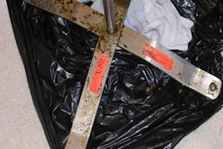
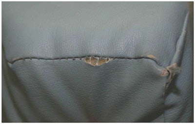
One much overlooked concern is the torn and frayed pads used in imaging departments and centers. Once the covering material has been breached, pads cannot be properly cleaned and should be immediately removed and replaced. This was clearly demonstrated by Shigeharu Oie, Ph.D., and colleagues from Yamaguchi University Hospital in Ube, Japan. In their article, "Contamination of Environmental Surfaces by Staphylococcus aureus in a Dermatological Ward and Its Preventive Measures," they stated, "Items with a smooth surface can be repeatedly used without problems if disinfected. However, on items with a porous surface made of a spongy material, S. aureus was detected even after disinfection had been done. Thus, porous surfaces made of such material cannot be adequately disinfected" (Biological and Pharmaceutical Bulletin, 2005, Vol. 28:1, pp. 120-123).
In the late 1980s and early 1990s when many of the pad systems in use today were developed, they were not designed to take the wear and tear of 5,000 to 10,000 patients a year for so many years. The fabric covers were coated with some type of plastic to make it waterproof. However, this plastic wears off, especially with cleaning solutions and with use. As a result, pad coverings have worn out, exposing the foam core, or have lost their ability to prevent penetration of bacteria and fluids into the central core, where it is impossible to clean.
 |
| Torn and frayed table pad. Image courtesy of Dr. Peter A. Rothschild. |
Only in the last five to10 years have hospital-acquired infections become so significant. Before that time, there was very little concern for contamination, and MRSA was not as prevalent as today. Therefore, pads on most imaging tables do not incorporate newer technologies developed to assist in infection control. Permanent antimicrobial agents should be incorporated into all table pads and positioners, as well as scanner controls, keyboards, etc. For added protection, the seams of the table pads should not only be tightly sewn, but also welded closed or have another permanent barrier in place in addition to simple stitching. The integrity of these seams is crucial in protecting patients.
Another area of concern is that of aerosolization of MRSA bacteria. Table pads inherently have air within them. When a patient lies down on the pads, this air is forced out through any hole or seam in the covering materials. This can cause the bacteria contaminating the central foam core to become ejected from the pad and aerosolized into the room environment. Of course, the reverse airflow caused by the patient rising off the pads causes infectious materials to be drawn into the foam core from the surface, which is then re-ejected into the air when the next patient lies on the pad.
Numerous articles have discussed the possibility of MRSA or other pathologic microorganisms becoming airborne during activities such as bed making, thus the possibility that MRSA can be transmitted among patients through the air. There also is a suggestion that airborne MRSA may play a role in MRSA colonization of the nasal cavity or respiratory tract. Research has shown that the presence of airborne MRSA in an area is strongly related to the presence and number of MRSA colonies and infected patients in that area. In a 2001 study on the airborne transmission of MRSA in a hospital environment, the researchers found that MRSA was recirculated among the patients, the air, and objects such as sinks, floors, and bed sheets. They concluded that measures should be taken to prevent the spread of airborne MRSA to control nosocomial MRSA infection.
This is clearly another reason why all pads must be inspected with a magnifying glass, and if any holes or loss of integrity of the covering material in any way is detected, the pads must be replaced.
Black light detection
Periodically testing all pads using a black light to detect biological contamination is also important. A black light provides light in the ultraviolet wavelengths that are especially sensitive in detecting biological material such as blood, fingerprints, and body fluids. Biological material remaining on the pads will light up under black light exposure. This is an excellent way to confirm that cleaning procedures are adequate.
If biological material remains after proper cleaning, this may indicate the covering material has been frayed or breached, thus allowing fluids to seep into the fabric itself and possibly penetrate to the underlying foam. Experiments performed by Ryan Whitney, a medical student who experimented with MRSA and MRI pads, have shown that MRSA can even go through frayed fabric covers without a tear and enter the central core, which is impossible to disinfect. This also demonstrates the need for pads and positioners to contain permanent antimicrobial agents in the covers and foam cores. More studies are needed to more thoroughly understand the depth of this problem and exactly how many pads in use today are contaminated.
A product called Glo Germ, which contains plastic "simulated germs," can be used for hand-washing training. Glo Germ liquid is rubbed on hands like lotion. For surface cleaning, Glo Germ powder is dusted onto surfaces and generally throughout the entire area. Hands are then washed or areas cleaned as normal. Under normal light, hands and surfaces will appear clean. However, ultraviolet light will show any "Glo Germs" remaining. This is an excellent way to train personnel in proper procedures for hand washing and pad cleaning.
The magnet bore
An area of proven risk for MRSA is the inside of the MRI unit itself, often referred to as the magnet bore or tunnel. The risk of MRSA transmission is increased in this area because the patient is often touching or in close contact with the bore surface. Cleaning inside the bore of an MRI unit can be a difficult, dangerous, and cumbersome task. The fact that most cleaning tools cannot even be brought inside the MRI room, and especially into the bore, makes this task even more difficult.
The best possible way to clean the bore is to physically crawl inside to clean and disinfect the entire bore by hand. Unfortunately, this also puts the technologist in close contact with the contaminated surfaces and is yet another reason this is almost never done. In fact, in over 25 years, I have never seen a cleaning crew or technologist clean the inside of the MRI bore.
One alternative for cleaning, sanitizing, and disinfecting inside the MRI bore and its surroundings is to use a cleaning tool long enough to reach well inside the bore with some kind of pad or sponge at its end soaked with a disinfectant.
MagnaWand has invented such a cleaning tool that is completely nonmagnetic and designed exclusively to clean the MRI bore and its surroundings. With the MagnaWand tool, technologists can easily reach inside the bore and clean surfaces using a disinfectant. Once the area of interest has been cleaned and disinfected, the disposable pad is simply ejected from the tool without being touched by the technologist's hands, eliminating the risk of further contamination.
By Dr. Peter A. Rothschild
AuntMinnie.com contributing writer
June 27, 2008
Part III of the series will discuss how the current state of MRI practice management contributes to poor infection control policies.
Dr. Rothschild is co-founder and medical director of High Field & Open MRI of Louisville, KY. He also is the president and founder of Patient Comfort Systems and Image Enhancement Systems, both in Hayward, CA. Since 1988, Rothschild has practiced exclusively in the area of MRI. He helped develop the first commercially available open MRI scanner while at the University of California, San Francisco, and pioneered the clinical aspects of open MRI, edited the first medical textbook on the subject, and has authored numerous medical journal articles about various aspects of MRI.
Related Reading
Sink or swim: Why a hand sink is crucial in the MR suite, March 14, 2008
Joint Commission MR safety surveys: Moving past the fire extinguisher, August 29, 2007
MR guidelines: Elevating standards of practice and care, March 10, 2007
How to calculate your MRI suite safety score, February 8, 2007
References
Blythe D, Keenlyside D, Dawson SJ, Galloway A. Environmental contamination due to methicillin-resistant Staphylococcus aureus (MRSA). J Hosp Infect. 1998;38(1):67-69. 1998;39(3):243-244.
Boyce JM, Pittet D. Guideline for hand hygiene in health-care settings. MMWR Morb Mortal Wkly Rep. 2002;51(16):1-44.
Boyce JM, Potter-Bynoe G, Chenevert C, King T. Environmental contamination due to methicillin-resistant Staphylococcus aureus: Possible infection control implications. Infect Control Hosp Epidemiol. 1997;18(9):622-627.
Cosgrove SE, Qi Y, Kaye KS, Harbarth S, Karchmer AW, Carmeli Y. The impact of methicillin resistance in Staphylococcus aureus bacteremia on patient outcomes: Mortality, length of stay, and hospital charges. Infect Control Hosp Epidemiol. 2005;26(2):166-174.
Delaney LR, Gunderman RB. Hand hygiene. Radiology. 2008;246(1)15-19.
Engemann JJ, Carmeli Y, Cosgrove SE. Adverse clinical and economic outcomes attributable to methicillin resistance among patients with Staphylococcus aureus surgical site infection. Clin Infect Dis. 2003;36(5):592-598.
Guidelines for Environmental Infection Control in Health-Care Facilities: Recommendations of CDC and Healthcare Infection Control Practices Advisory Committee (HICPAC). Centers for Disease Control and Prevention Web site. http://www.cdc.gov/ncidod/dhqp/gl_environinfection.html. June 2003.
Huang R, Mehta S, Weed D, Price CS. Methicillin-resistant Staphylococcus aureus survival on hospital fomites. Infect Control Hosp Epidemiol. 2006;27(11):1267-1269.
Huang SS, Datta R, Platt R. Risk of acquiring antibiotic-resistant bacteria from prior room occupants. Arch Intern Med. 2006;166(18):1945-1951.
Kanal E, Barkovich AJ, Bell C, et al. ACR Guidance Document for Safe MR Practices: 2007. AJR. 2007;188(6):1447-1474.
Kerr KG, Beggs CB, Dean SG, et al. Air ionisation and colonisation/infection with methicillin-resistant Staphylococcus aureus and Acinetobacter species in an intensive care unit. Intensive Care Med. 2006;32(2):315-317. 2006;32(9):1439.
Kihlstrom J. Hand Washing by Health Care Providers. Institute for the Study of Healthcare Organizations & Transactions Web site. http://www.institute-shot.com/hand_washing_by_health_care_providers.htm. 2000.
Lankford MG, Collins S, Youngberg L, et al. Assessment of materials commonly used in health care: Implications for survival and transmission. Am J Infect Control. 2006;34(5)258-263.
Martinez JA, Ruthazer R, Hansjosten K, Barefoot L, Snydman DR. Role of environmental contamination as a risk factor for acquisition of vancomycin-resistant enterococci in patients treated in a medical intensive care unit. Arch Intern Med. 2003;163(16):1905-1912.
Oie S, Yanagi C, Matsui H, Nishida T, Tomita M, Kamiya A. Contamination of environmental surfaces by Staphylococcus aureus in a dermatological ward and its preventive measures. Biol Pharm Bull. 2005;28(1):120-123.
Scanlon T, Murray J. MRSA detection in the radiology department. Ir J Med Sci. 2005;174(4),es3:10.
Shiomori T, Miyamoto H, Makishima K. Significance of airborne transmission of methicillin-resistant Staphylococcus aureus in an otolaryngology-head and neck surgery unit. Arch Otolaryngol Head Neck Surg. 2001;127(6):644-648.
Shiomori T, Miyamoto H, Makishima K, et al. Evaluation of bedmaking-related airborne and surface methicillin-resistant Staphylococcus aureus contamination. J Hosp Infect. 2002;50(1):30-35.
Wilson RD, Huang SJ, McLean AS. The correlation between airborne methicillin-resistant Staphylococcus aureus with the presence of MRSA colonized patients in a general intensive care unit. Anaesth Intensive Care. 2004;32(2):202-209.
Copyright © 2008 Dr. Peter Rothschild