FDG-PET/CT should be used in conjuction with breast MRI for patients with locally advanced breast cancer, according to researchers from Stanford University Medical Center in Stanford, CA.
The hybrid imaging modality should serve as a complementary imaging tool because it can detect distant metastases outside the field-of-view of breast MRI, they explained.
The researchers examined breast cancer patients using the two imaging modalities. They found that PET/CT examinations alone identified 11 women, or 23.4%, of 47 patients with distant metastases who had breast MRI and PET/CT examinations performed at approximately the same time.
The 47 patients were a subgroup of a larger retrospective study of 181 consecutive women who had FDG-PET/CT scans performed for initial staging (20 patients) or restaging (161 patients) over a four-year period from 2003 to 2006. The research team reviewed and made independent diagnoses of the 181 PET/CT exams and 47 breast MRIs to assess the sensitivity and specificity of PET/CT by itself and also compared with breast MRI.
Sensitivity and specificity were calculated using pathology results or clinical follow-up as the gold standard. Tumor size at cancer presentation ranged from 0.9 to 7.5 cm (average 2.68 ± 1.6 cm).
As a single exam, FDG-PET/CT had 92.9% sensitivity and 90% specificity for distant metastases, according to Dr. Andrei Iagaru, who presented the study findings at the 2007 RSNA meeting in Chicago. For breast disease, the modality had a sensitivity of 81.5% and a specificity of 98.4%. While PET/CT had excellent specificity at 97.9%, its sensitivity for axillary staging was limited at 76.9%.
 |
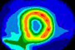
| A 42-year-old woman with infiltrating ductal carcinoma. Postoperative (14 days) PET/CT demonstrated liver metastases (arrows). All images courtesy of Stanford University Medical Center. |
Breast MRIs of the 47 patients were 5.2% more sensitive than whole-body PET/CT performed at the same time (87.5%, compared with 83.3%). The specificity of breast MRI was similar to PET/CT at 93.3%, compared to 94.1% for PET/CT.
Iagaru noted that this study supports the results of a similar research study that he participated in at the PET Imaging Science Center at the University of Southern California (USC) in Los Angeles. "Over a three-year period from June 2002 to May 2005, 21 patients with breast cancer had breast MRI and whole-body FDG-PET/CT examinations performed over an interval of two to188 days," he added.
The results of the retrospective study evaluating these examinations were published in the Annals of Nuclear Medicine (2007, Vol. 21:1, 33-38).
In the USC study, six patients had procedures performed in the preoperative period. Breast MRI identified breast lesions in four patients; PET/CT identified breast lesions, which were malignant on pathology examination, in five patients.
 |
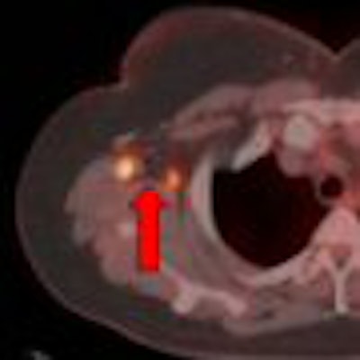
| Image of a 47-year-old woman with breast carcinoma shows how PET/CT demonstrated multifocal right breast disease (horizontal arrows) and right axillary lymph nodes metastases (vertical arrows). |
Fifteen patients had the procedures performed four to 175 days after surgery. The PET/CT revealed metastatic disease in six patients with 100% sensitivity and 90% specificity. "As a result of these findings," Iagaru said, "the staging and the management of the disease for these patients were altered."
The role of PET/CT in addition to breast MRI is not completely elucidated, he added. That is the primary reason that the two studies were performed. "Both of these studies suggest that FDG-PET/CT and breast MRI should be considered as complementary imaging tools for the staging of breast cancer patients with large tumors, aggressive histology, or equivocal findings from other imaging studies."
By Cynthia Keen
AuntMinnie.com contributing writer
April 4, 2008
Related Reading
PET/CT beats 3T MRI in whole-body primary tumor staging, March 10, 2008
FDG-PET/CT aids in radiation therapy planning, March 5, 2008
PET/CT moves closer to diagnostic standard of care, March 1, 2008
False positives a concern with FDG-PET/CT for head, neck tumors, January 31, 2008
PET/CT shows its worth in cervical carcinoma, January 18, 2008
Copyright © 2008 AuntMinnie.com