SEATTLE - Before the first image is even taken, radiologists conducting MR studies of the liver must have a thorough knowledge of a patient's clinical history, according to a presentation Saturday at the International Society for Magnetic Resonance in Medicine (ISMRM) meeting. This information will help determine the category of liver disease the patient may have, which is crucial for guiding the imaging exam.
"Whenever I see any patient, I like to have as much clinical history up front before I make the diagnosis, and then afterward evaluate the MRI findings," said Dr. Richard Semelka from the University of North Carolina in Chapel Hill. "There are basically three categories of patients: those with no known disease, those with primary malignancy, and those with chronic liver disease."
The patient's clinical history is important "because the likelihood of having any particular lesion is very much dependent on what category the patient falls into," added Semelka, who is a consultant for Bracco Diagnostics of Princeton, NJ.
No known disease
About 20% of the general population has benign liver lesions, according to Semelka. In fact, based on actuarial data, benign lesions are 10 to 100 times more common than malignant ones, he said. So if malignant disease is that rare, radiologists need to look elsewhere in cases of unknown disease, he stressed. Questions that need to be answered include the following:
- What does normal background of the liver look like?
- Could the patient have cirrhosis?
- Despite the odds, could the patient have an unsuspected primary malignancy?
Semelka listed a number of alternate diagnoses to cancer, such as biliary hamartoma, hemangioma, and foregut cyst. In the case of the latter, Semelka warned that cysts may have an enhancing capsule on MR, which is also a hallmark feature of metastases.
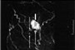
MRI features of hemangioma include high signal intensity, slow nodular enhancement, distinctive retention of contrast, and no washout. "There's nothing that comes close to the accuracy of MR to diagnose the full range of hemangioma," Semelka said. "I like to describe hemangiomas as nodules that enlarge and coalesce."
Finally, there is adenoma, characterized by small, incidental lesions that are either isointense or hypointense. Semelka said that in his reports, he prefers to qualify how bright dark lesions are by describing them as minimally, moderately, or markedly intense.
Unexpected primary malignancy
This category can include islet cell tumor metastasis, which is not bright on T2-weighted MRI and generally does not cause ductal obstruction.
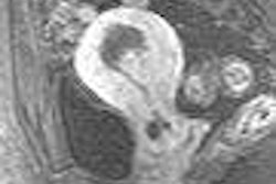
In cases of colon cancer with liver metastases, look for archiform enhancement and architecture, Semelka said. The cauliflower appearance of the liver lesion is a distinctive, histological feature of metastases with colon cancer as the primary tumor.
"The critical questions are: What is the histopathological type and location of the primary tumor? Was chemotherapy administered and when? Acute response to chemotherapy is very different from chronic response and it's important to know that," Semelka said, adding that liver metastases can adopt various appearances depending on the time and duration of treatment.
Chronic liver disease
Again, Semelka pointed out that the occurrence of carcinoma in the general population is low enough that imaging experts must consider chronic liver disease -- even if the MRI reveals features that are commonly associated with carcinoma.
Chronic liver disease is marked by mild hyperintensity on T2-weighted imaging, washout with late capsule enhancement (a hallmark of metastases, he reiterated), and early intensity enhancement.
Other possible diagnoses are listed below:
- Regenerative nodules (dark lesions usually indicative of iron)
- Low-grade dysplastic nodues (larger than background liver)
- High-grade dysplastic nodules (require close follow-up to see if underlying liver disease exists)
- Fatty hepatocellular carcinoma (HCC, high signal on T1-weighted imaging, calls for in-phase and out-of-phase imaging)
- Hypovascular HCC
For hypovascular HCC, if the MR exam is not being done with CT- or MR-guided hepatic arterial injection, then readers should pay close attention to the late postcontrast images before making the diagnosis, Semelka said.
MRI pearls
In terms of sequences, Semelka endorsed T1-weighted imaging with in-phase and out-of-phase imaging, and T2-weighted imaging with and without fat suppression and hepatic arterial-dominant phase enhancement. He stressed that the latter is most critical for making the differential diagnosis. Semelka also recommended that if portal venous and interstitial phase-enhancement sequences are done, then one full set of each must be completed.
Semelka wrapped up his talk with some take-home points:
- Hemangioma is always bright on T2-weighted imaging.
- HCC is never bright on T2-weighted imaging.
- Is the liver cirrhotic? Could it be HCC? Look for fibrosis on short TE, T1-weighted images.
- Could the lesion be related to chemotherapy?
By Shalmali Pal
AuntMinnie.com staff writer
May 7, 2006
Related Reading
Targeted angiography precludes unintentional embolization during TACE, May 28, 2006
Detection of liver metastases with contrast-enhanced ultrasound, March 23, 2006
MRI after CT cuts need for liver biopsies in cancer patients, May 19, 2005
Breath-hold technique comparable to respiratory-triggered MR for liver imaging, March 16, 2005
Exam technique critical in liver ultrasound, October 22, 2004
Copyright © 2006 AuntMinnie.com