Three-dimensional contrast-enhanced MR angiography (CEMRA) at 3 tesla is "extremely promising" for evaluating abdominal vasculature, according to researchers from the University of California, Los Angeles. The advantages of 3D CEMRA at 3 tesla include improved spatial resolution and a higher signal-to-noise ratio (SNR).
Dr. Kambiz Nael, a research fellow at UCLA, and his colleagues used this MR technique to assess renal and visceral arterial branches in 25 patients with suspected arterial stenosis. A whole-body 3-tesla system with an 8-channel body-array coil was employed. The patients received 25 mL of gadodiamide at 1.5 mL/sec. A fast 3D GRE sequence was used with images acquired in the coronal plane during a 20-second breath-hold.
The patients first practiced the 20-second breath-hold before undergoing the imaging, Nael explained in a talk at the 2005 RSNA meeting in Chicago. None of those patients had difficulty with holding their breath for the duration of the scan, he said.
Two radiologists reviewed 13 arterial segments, including the bilateral renals and the celiac trunk, in order to assign a score between 1 (insufficient for diagnosis) and 5 (excellent). Vascular occlusive disease was scored based on a three-point scale, with 3 representing occlusion. At the time Nael presented the study, six patients' had their MRA results confirmed with conventional angiography.
 |
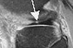
| Coronal 3D volume rendered from CEMRA in a 54-year-old man with uncontrolled hypertension. By acquiring images over a large field-of-view on a high-resolution matrix, the arteries of the entire abdomen, pelvis, and lower thorax are visualized in detail. |
According to the results, all 13 segments in all subjects were visualized. There were 54 segments with vascular pathology, including seven with segmental occlusions. Renal artery stenosis was detected in 18 segments (12 patients), including 13 segments with more than 50% occlusion. In six patients, MRA results were confirmed with angiography, giving MRA a sensitivity and specificity of 100% for evaluating renal artery stenosis, Nael stated.
 |
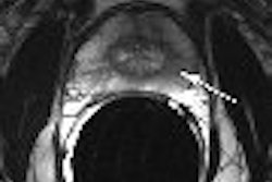
| Targeted MIP (20 mm) from same patient as above. By applying parallel acquisition with an acceleration factor of 3, the 3D dataset was acquired with 0.8 x 0.7 x 0.9 mm³ voxels over a 420-mm field-of-view, during a 19-second breath-hold. Note the branch stenosis at the origin of the left inferior pole renal artery. |
In addition to higher SNR, using a parallel acquisition at 3 tesla can improve spatial resolution and generate 0.50 mm3 voxels, Nael's group concluded. This MR technique may well rival the spatial resolution of multislice CT, they added.
 |
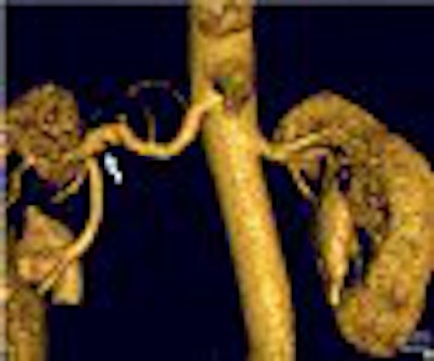
| Coronal oblique 3D volume rendered (above) and thin MIP (below) from CEMRA, in a 60-year-old woman with uncontrolled hypertension, show beaded irregularity involving the distal right renal artery (arrows) consistent with fibromuscular dysplasia (FMD). Images courtesy of Dr. Kambiz Nael. |
 |
Nael told AuntMinnie.com that 3-tesla MRA is the first choice for evaluating abdominal arteries in his institution, while CT angiography is reserved for cases with MRA contraindications.
An RSNA attendee said that when he performed 3D CEMRA at 3 tesla, artifacts at the lumen border appeared. He said he needed to use fat saturation to get rid of those artifacts.
Nael said his group did not have this problem and did not use fat saturation. He attributed the lack of lumen border artifacts to the short TE time of about 1.2 msec.
Nael's group published a more detailed description of their 3D CEMRA techniques in Magnetic Resonance Imaging Clinics of North America last year. Their review article covered aneurysms, dissections, occlusions, congenital lesions, and anatomic anomalies (May 2005, Vol. 13:2, pp. 359-380).
Nael also told AuntMinnie.com that the next phase of research will focus on continuing to improve spatial resolution by applying modified pulse sequences and more aggressive parallel acquisition techniques. They plan to share their results in an upcoming issue of the American Journal of Roentgenology.
"Our experience to date suggests that CEMRA at 3 tesla is robust and frequently yields spectacular images using lower contrast doses than are typical at 1.5 tesla," Nael said. "We now use 3 tesla routinely for CEMRA of the carotids, thorax, abdomen, and pelvis, and have found, so far, that patient acceptance is similar at 1.5 tesla and 3 tesla."
By Shalmali Pal
AuntMinnie.com staff writer
January 20, 2006
Related Reading
Studies showcase MRA at its best in arteries and combined with CAD, November 17, 2005
3-tesla scanning fails to improve coronary MRA's accuracy, November 11, 2005
Copyright © 2006 AuntMinnie.com