Pediatric PET/CT imaging times can be reduced by 30% for many patients without a loss of diagnostic utility, according to researchers from Seattle Children's Hospital and the University of Washington Medical Center in Seattle.
The researchers found, however, that their data did not support reduced acquisition times or lowered FDG doses for patients weighing less than 22 kg (48.4 lb). Lead study author Marla Sammer, MD, presented the findings at last week's annual SNM meeting in Salt Lake City. The research received SNM's Pediatrics Council Young Investigator Award.
Because FDG dose and acquisition times for pediatric patients generally are based on adult guidelines, the researchers sought to determine if shorter acquisition times or a lower FDG dose could be used for pediatric FDG-PET/CT exams while maintaining diagnostic image quality.
"Reduction of overall scan time potentially reduces motion artifacts, improves patient comfort, and decreases length of sedation," the authors wrote. "Alternatively, decreased FDG dose minimizes radiation risk."
Fourteen patients were included in the study, with one patient scanned twice. The patients ranged in age from 1 to 23 years, with a mean age of 8.8 years. There were nine males and five females; weights ranged from 13 kg (28.6 lb) to 109 kg (240 lb). Three patients weighed less than 22 kg, 11 patients weighted more than 22 kg, and two patients weighed more than 70 kg.
The FDG injected dose was based on 0.144 mCi/kg, with fixed acquisition times of three minutes for patients weighing less than 22 kg and five minutes for those weighing more than 22 kg.
Seven of the 14 exams were to determine cancer staging, while the other seven were performed after treatment. The diseases included four cases of non-Hodgkin's lymphoma, two cases of Ewing's sarcoma, and two cases of neuroblastoma. In addition, there was one case each of Hodgkin's lymphoma, malignant fibrous histiocytoma, osteosarcoma, hepatoblastoma, and Castleman's disease.
The radiologists then assessed the results to determine if the PET/CT scan was adequate for evaluation and to quantify the number of lesions. The whole-body PET/CT scan was divided by body region by the neck, chest, abdomen, pelvis, and bones, and the number of lesions was counted in each area for both an individual and total number of lesions.
For each patient, multiple datasets with image acquisition times ranging from one minute to five minutes were used to obtain the field-of-view. Fifty-six image volumes were generated, randomized, and reviewed blindly with corresponding CT image volumes by five radiologists. Both overall adequacy and lesion detection accuracy and confidence by body region were evaluated.
"In patients who are greater than 22 kg, our standard acquisition time is five minutes," Sammer said in her presentation. The radiologists "would automatically shorten the data into its lower time points, usually for three, two, and one minute, and [the data] would be stored."
For patients weighing 22 kg or less, the standard acquisition time of three minutes was shortened to two- and one-minute time points, she added.
The analysis showed that the average threshold of acquisition time for an adequate PET/CT image for patients weighing less than 22 kg was 2.3 minutes. In patients weighing between 22 and 70 kg, the average threshold of acquisition time was two minutes, and for patients weighing more than 70 kg, the time was three minutes.
Detection accuracy
Lesion detection accuracy and diagnostic confidence was not degraded from five minutes to one minute for the field-of-view for abdominal lesions, but chest lesion detection became less accurate when imaging acquisition was reduced more than 30%.
 |
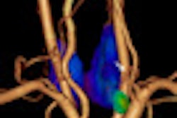
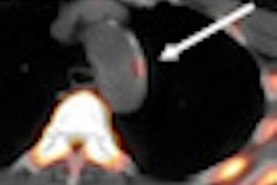
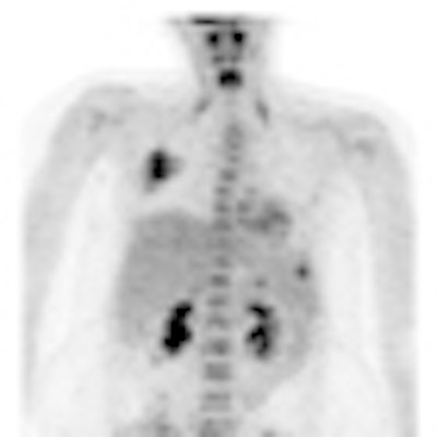
| PET/CT scan of an 18-year-old, 94 kg (207 lb) female patient who was imaged for the staging of osteosarcoma. The five-minute image acquisition time was considered adequate, as was the image obtained at three minutes. Images acquired at two minutes and one minute were deemed inadequate. However, lesion detection was degraded at one minute and was preserved at two minutes and longer. The adequate field-of-view for this patient was three minutes, so dose potentially could have been reduced by 40% to 60%. Image and information courtesy of Marla Sammer, MD. |
Although images generated from shorter acquisition times suggest that imaging duration can be reduced by 30% without a loss of diagnostic image quality in pediatric PET/CT, that wasn't the case for patients weighing less than 22 kg, the researchers concluded.
"We have only three patients [weighing less than 22 kg], so, before we have any further conclusions, we need to evaluate more patients in more scans," Sammer said. "When the patients were less than 70 kg, we were all comfortable with exams at 40% [reduced] time and, hopefully, dose reduction."
While lesion detection was achieved with a 20% reduction in image acquisition time, she added that it was "relatively difficult to remain consistent about the number of lesions detected between the time points, but we can say that below 60% or greater reduction [in image acquisition time], we did not preserve the number of lesions detected."
By Wayne Forrest
AuntMinnie.com staff writer
June 17, 2010
Related Reading
Image Gently expands dose awareness to nuclear medicine, June 3, 2010
Dose can be reduced by 75% in pediatric chest CT exams, June 18, 2009
Color-coded CT protocols help reduce pediatric radiation dose, June 4, 2009
SPR news: Rads must take lead in reducing pediatric CT dose, April 23, 2009
ARRS study: Child's body shape can reduce CT dose, April 23, 2009
Copyright © 2010 AuntMinnie.com