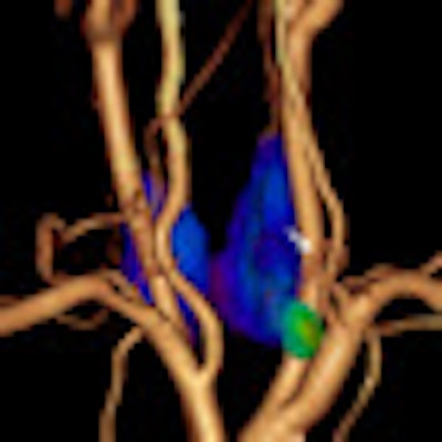
The annual SNM Image of the Year for 2010 comes from researchers at Hokko Memorial Hospital in Sapporo, Japan, who fused images of MDCT and technetium-99m sestamibi (Tc-99m MIBI) SPECT to provide information regarding detailed anatomical localization, angiography, and metabolism of an enlarged parathyroid gland.
The study, titled "Clinical value of fusion images of MIBI SPECT and enhanced MDCT registered by workstation in primary hyperparathyroidism," was led by Kunihiro Nakada, MD. The image was chosen from more than 600 abstract presentations and some 750 scientific posters as an example of simultaneous multimodality imaging.
The prospective study included 31 patients with biochemical evidence of primary hyperparathyroidism who were scanned by MIBI SPECT and CT on the same day. Early (20 minutes postintravenous injection) and delayed (120 minutes postinjection) planar and SPECT images of Tc-99m MIBI were acquired using a dual-head gamma camera.
Thin-slice (2-mm) multiplanar reconstruction images of the neck also were obtained using 64-detector-row MDCT with contrast enhancement. Patients also received neck ultrasound.
When researchers discovered an enlarged gland, volume-rendered images of the thyroid and parathyroid were generated.
Fused images
Finally, 2D and 3D fusion images of MIBI SPECT and MDCT were obtained using dedicated workstations to compare the diagnostic value of SPECT/CT fusion images with MIBI SPECT alone and by ultrasound.
The study detected a total of 34 enlarged glands that also were identified by surgery. SPECT/CT fusion imaging identified 32 (94%) of 34 enlarged glands, MIBI SPECT found 27 (79%) enlarged glands, and ultrasound discovered 27 (79%) enlarged glands.
Five glands missed by ultrasound or planar MIBI scintigraphy were additionally localized by fusion images. In addition, lymph nodes adjacent to the thyroid were accurately distinguished from enlarged parathyroid gland in four patients.
With use of the 2D and 3D fusion images, "preliminary results in eight patients showed that average operation time is decreased to about 82% of that without fusion images," the researchers noted.
 |
| SNM's Image of the Year is an example of simultaneous multimodality imaging. A parathyroid adenoma (green) is identified on MIBI SPECT, while blood vessels are seen on a CT angiogram performed during the same acquisition on a SPECT/CT camera. Image courtesy of SNM. |
"Fusion images of MDCT and MIBI SPECT provide highly helpful information regarding detailed anatomical localization, angiography, and metabolism of enlarged parathyroid gland," the researchers concluded.
For the first time in 53 years, the SNM Image of the Year was not chosen by now-retired nuclear medicine patriarch Henry Wagner, MD, who did not attend the annual meeting. This year's image was selected by a committee of SNM members.
By Wayne Forrest
AuntMinnie.com staff writer
June 14, 2010
Related Reading
SNM: 1 year later, Mo-99 shortage 'a lot worse,' June 8, 2010
Novel agents and new tools chart molecular imaging's future, June 8, 2010
SNM meeting opens with call for effectiveness research, June 7, 2010
SNM convenes annual meeting as challenges persist, June 3, 2010
Copyright © 2010 AuntMinnie.com