Researchers in Japan have found that uptake of FDG in PET/CT scans of acute aortic dissection is significantly associated with an increased risk of rupture and poor patient outcomes, according to a new study published in the May issue of the Journal of Nuclear Medicine.
The researchers also concluded that FDG-PET/CT may be used to better manage patients with acute aortic dissection (AAD), but more studies are needed to determine the modality's role in this clinical application. The lead study author is Dr. Kimihiko Kato from the department of cardiovascular medicine at Gifu Prefectural Tajimi Hospital in Tajimi (JNM, May 2010, Vol. 51:5, pp. 674-671).
Seventeen men and 11 women with acute aortic dissection were enrolled in the prospective study at the hospital from April 2006 to November 2008. The study also examined 14 age- and sex-matched patients as a control group.
After initially receiving a chest x-ray, all patients also were given an electrocardiogram, an echocardiogram, and a contrast-enhanced CT scan. A series of 64-slice CT exams (Aquilion 64, Toshiba Medical Systems, Otawara, Japan) also were given to evaluate progression, rupture, and morphologic parameters, such as maximum aortic dissection diameter, diameter of true and false lumen, and the presence of intramural hematoma.
In addition, patients received unenhanced CT and PET images acquired consecutively at 50 and 100 minutes after injection of 4 to 6 MBq/kg of FDG, using a PET/CT system (Biograph 6, Siemens Healthcare, Erlangen, Germany).
Of the 28 AAD patients, eight patients experienced an unfavorable outcome, with two dying from rupture, four patients required surgery, and two patients progressing to acute aortic dissection. The remaining 20 patients had favorable acute aortic dissection outcomes.
 |
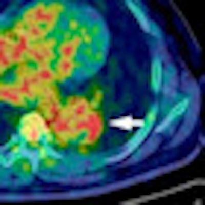
| Above: Enhanced CT (left) shows acute aortic dissection consistent with low uptake of FDG in intramural hematoma lesion on transaxial PET/CT at 50 minutes (middle) and 100 minutes (right). This 82-year-old woman experienced acute aortic dissection regression two months after onset but had a favorable outcome. Below: Enhanced CT (left) shows acute aortic dissection consistent with high uptake of FDG in dissected aortic wall on transaxial PET/CT at 50 minutes (middle) and 100 minutes (right). This 44-year-old man underwent elective repair of aortic aneurysm because of acute aortic dissection progression three months after onset and experienced an unfavorable outcome. Images courtesy of JNM. |
 |
The researchers found that standardized uptake values (SUV) of FDG on PET were greater in AAD patients than in controls. In addition, among the AAD patients, those patients with unfavorable outcomes had greater SUV at the site of maximum aortic dissection than those with favorable outcomes.
A mean SUV greater than 3.029 has significant predictive power, according to the authors, with sensitivity of 75.0%, specificity of 70.0%, positive predictive value of 50.0%, negative predictive value of 87.5%, and accuracy of 71.4%.
In a perspective article in JNM on the role of FDG-PET in aortic dissection, Dr. James H.F. Rudd, Ph.D., a researcher and consultant cardiologist at the University of Cambridge in the U.K., wrote, "Early diagnosis and treatment are essential for survival of patients with this rare and often fatal disease. Although further studies are needed, this research suggests that FDG-PET imaging might be used to identify patients who are at a very high risk of complications, allowing them to be fast-tracked to surgery."
By Wayne Forrest
AuntMinnie.com staff writer
April 30, 2010
Related Reading
MDCT highly sensitive for aortic dissection, March 29, 2006
Copyright © 2010 AuntMinnie.com