Researchers at the Cleveland Clinic in Ohio say physicians should consider a second FDG-PET scan to help localize seizure origin in patients with refractory epilepsy whose initial images show regional hypermetabolism.
The study, presented at the 2009 RSNA annual meeting, also suggests that it may be worthwhile to repeat the FDG-PET exam if no regions of hypometabolism are found, but an electroencephalogram (EEG) uncovers frequent spiking activity.
Epilepsy is a common neurological condition in approximately 1% of the population, and surgical treatment has proved promising for patients with refractory epilepsy. Previous research has found that regional hypometabolism is a strong predictor of a good surgical outcome.
"In this study, we investigated different kinds of pathologies in epilepsy on FDG-PET imaging," said lead author Dr. Roland Talanow. "We wanted to find out the different kinds of etiologies which cause refractory epilepsies."
Retrospective review
The study retrospectively reviewed FDG-PET images of 280 patients (135 females and 154 males) with refractory epilepsy. The patients, who ranged in age from 1 day to 64 years, also were treated surgically for epilepsy.
All patients received an FDG-PET exam (ECAT HR+, Siemens Healthcare, Malvern, PA) for their presurgical workup. Patient preparation included fasting for at least four hours, no insulin injections within six hours before the study, and EEG monitoring 30 minutes prior to and 40 minutes after FDG injection. Patients also were seizure-free for at least 24 hours before the FDG-PET exam.
Metabolism levels were defined as hypermetabolic, hypometabolic, or normal. The region of abnormal metabolism was evaluated visually and semiquantitatively and compared to the contralateral side of the same region. The FDG-PET images were then compared to the surgical pathology reports.
Pathology findings
In the analysis, the researchers found that the majority of pathologies were regional hypometabolism. FDG hypometabolism was demonstrated by 153 of 166 (92.1%) patients with sclerosis. In addition, nine of 166 patients (5.4%) had normal metabolism and four patients (2.4%) had increased FDG uptake (hypermetabolism).
Results also showed that 60 of 71 patients (84.5%) with mild cognitive disorder demonstrated hypometabolism, while six patients (8.4%) exhibited hypermetabolism and five patients (7%) showed normal metabolism. FDG-PET images also revealed regional hypometabolism in all of the neoplasms (24 of 24 patients) and inflammatory process (25 of 25 patients).
"FDG-PET has the advantage compared to CT and other anatomical modalities in that it gives functional information," Talanow said. "MRI has some limited ability to provide functional information, but so far no study has shown that it is equal to FDG-PET."
 |
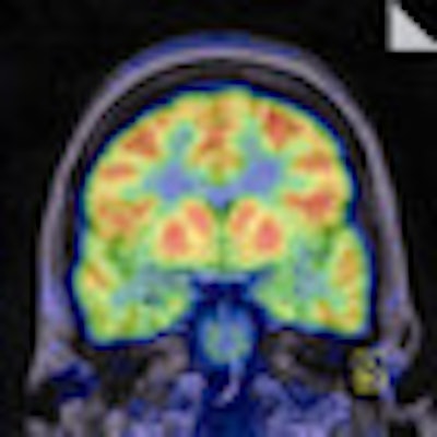
| A noncontrast T1-weighted MR image (left) shows fairly symmetric and moderate volume loss involving bilateral hippocampi, which illustrates a slight abnormal signal. The PET scan (right) is taken after an injection of 7.2 mCi FDG and shows symmetric FDG uptake in the cerebral cortex, subcortical gray matter, and cerebellum without definite focal area of hypometabolism. Image courtesy of Dr. Roland Talanow, the Cleveland Clinic. |
While it is critical to note that the majority of patients with refractory epilepsy are hypometabolic (previous research has shown that hypometabolic activity is an indicator of increased lifetime seizure episodes), the researchers concluded that physicians should not overlook patients with regional hypermetabolism, as it, too, might sufficiently identify the seizure origin.
"The most significant finding is that the vast majority of pathologies showed hypometabolism," Talanow said, "but with 2% of the cases showing hypermetabolism, it suggests to repeat the [FDG-PET] study, because the hypermetabolic focus is most likely still the focus of the seizure."
A follow-up FDG-PET scan also may be beneficial if no regions of hypometabolism are found and EEG shows frequent spiking activity.
By Wayne Forrest
AuntMinnie.com staff writer
December 29, 2009
Related Reading
FDG-PET benefits findings in children with refractory epilepsy, July 2, 2009
3-tesla MRI both challenges and rewards pediatric radiologists, December 2, 2008
3-tesla MRI superior to 1.5-tesla MRI in epilepsy evaluation, October 1, 2008
Study shows benefits of very delayed cerebral FDG-PET imaging, December 21, 2007
PET helps predict surgical outcome in refractory epilepsy patients, December 13, 2007
Copyright © 2009 AuntMinnie.com