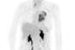
TORONTO - Cardiac images produced on gamma cameras in which patients are imaged in the sitting position may look different than those from standard supine-oriented gamma cameras, a phenomenon that clinicians should be aware of, according to a presentation at this week's SNM meeting.
Traditionally, SPECT myocardial perfusion imaging (MPI) exams have been conducted on gamma cameras with patients lying in the supine position. But a new generation of compact dedicated cardiac gamma cameras has hit the market in which scans are performed on sitting patients.
How could the different positions affect myocardial perfusion images? A group from the Center for Nuclear Medicine in Prague, Czech Republic, examined the issue by comparing images acquired with patients in both orientations, according to lead author Dr. Otto Lang.
The study population consisted of 31 patients with suspected or known ischemia who were imaged twice -- once on a dual-head camera with patients in the supine position (Spirit DH-V, Mediso Medical Imaging Systems, Budapest, Hungary) and again with patients on a sitting dual-head camera (CardioDesk, Mediso).
Upon analyzing the images, Lang and colleagues found that the heart appeared smaller and more vertical in sitting patients. Other quantitative details are shown in the following table:
Quantitative differences in MPI studies, supine versus sitting position
|
Total perfusion defect and mean summed stress score differed significantly. Analysis of individual vascular territories revealed only left anterior descending defect size to be significantly different, while defect sizes in the two other areas were similar.
Lang concluded by stating that the differences in quantitative values between supine and sitting positions were significant enough that sites using sitting cardiac cameras should not use normals databases derived from studies collected on patients in the supine position. "We should learn a new model or pattern of radioactive distribution," he said.
An SNM attendee in the audience commented that sites can develop new normals databases on their own by scanning several dozen patients at low probability for heart disease.
Related Reading
DIAD follow-up confirms SPECT MPI guidelines for diabetic patients, June 15, 2009
Hybrid coronary CTA, SPECT offer less radiation, shorter scan time, June 15, 2009
Gated SPECT best indicator for cardiac events, April 13, 2009
ACC study: Most cardiac SPECT scans are appropriate, April 1, 2009
MRI, MDCT err in estimating cardiac functional parameters, February 20, 2009
Copyright © 2009 AuntMinnie.com