TORONTO - Preliminary results of a new PET/MRI prototype breast imaging system show that the hybrid technology could produce images with enough clarity to more accurately classify lesions.
Researchers at Stony Brook University in New York are working with Dr. David Schlyer at Brookhaven National Laboratory (BNL) in Upton, NY, on the project. Bosky Ravindranath, research assistant and lead author of a study, presented details on the prototype's progress on Tuesday at the SNM annual meeting.
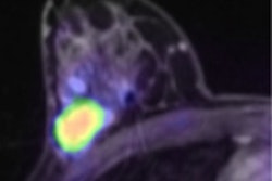
The motivation behind the project is to develop a high-resolution PET imaging system that can work inside an MRI scanner, combining MRI's high-resolution anatomic images with quantitative biochemical information from PET. "The combination of these two pieces of information will lead to better diagnosis, staging, and evaluation of breast cancer," Ravindranath said, "as well as research applications, such as radiotracers, MR imaging agents, and pulse sequences."
Prototype conception
The detector design for the PET/MRI prototype is based on a small animal imaging system developed at BNL called the Rat Conscious Animal PET, or RatCAP. The miniature PET scanner was designed to acquire 3D images of a rat's brain.
The prototype includes a modular 3D tomographic PET scanner that fits inside a dedicated breast MRI coil (Aurora Imaging Technology, North Andover, MA). "When the patient is positioned on the RF coil," she said, "the breast region to be imaged is surrounded by the PET system and is in the field-of-view of the RF coil as well. Therefore, we are able to obtain simultaneous PET/MR images."
The prototype has an inner diameter of 145 mm and an axial length of 96 mm. The design uses vertical stacks of lutetium orthosilicate (LSO) crystals arranged in a modular circle. By using this configuration, Ravindranath said there is "a possibility of expanding the field-of-view in order to accommodate a larger breast."
Phantom results
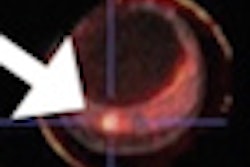
Using phantoms to test the prototype, Ravindranath said the results show that the researchers have obtained "good PET spatial resolution throughout the field-of-view using the prototype system." In addition, MRI tests with the PET insert also have shown good signal intensity and fat suppression capability.
"We have shown that PET can be operated unshielded inside the MRI, but we can improve the MR image quality by filtering the power supply cables to the MRI penetration panels," she added. "This way the RF shielding of the MRI room is maintained. That also has been reducing the noise and frequency artifacts."
The researchers plan next to begin working on the acquisition of simultaneous PET and MR images. Based on the preliminary results, they also soon expect to start testing the system on breast cancer patients.
The project is supported by the U.S. Department of Energy.
By Wayne Forrest
AuntMinnie.com staff writer
June 16, 2009
Related Reading
Open-source software delivers 3-way (PET/CT/MR) image fusion, April 25, 2008
Fused MRI and PET improve specificity of breast MRI, August 17, 2007
Aurora partners with Taiwan research center, March 2, 2009
Aurora wins breast MRI install in Italy, February 12, 2009
Aurora touts positive clinical study, November 10, 2008
Copyright © 2009 AuntMinnie.com