TORONTO - A new PET/MRI system can successfully provide whole-body images of rats and other small animals, potentially improving imaging capabilities and advancing research in many areas, according to a new study presented Sunday at the SNM annual meeting.
Lead study author Mitsuaki Tatsumi, an associate professor of radiology at Osaka University Graduate School of Medicine and Medical Hospital in Japan, presented the findings.
The results could open new opportunities in research in terms of increased understanding of kinetics, biodistribution of radiopharmaceuticals, and, eventually, disease characteristics and physiological processes, Tatsumi said. The next steps include imaging body tumors in rats or mice, and developing PET/MRI systems that can image people.
A combined PET/MRI system simultaneously delivers specific molecular information related to cell surface reactors, enzymes, and gene expression that PET provides, as well as anatomical data, soft-tissue contrast, and information about perfusion and permeability shown in MRI.
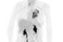
In the study, researchers injected rats with the radionuclide C-11 methionine for PET imaging and gadoxetate for contrast MRI images. Simultaneous PET and MR imaging was performed from head to abdomen. PET, MRI, and coregistered fusion images were then evaluated for image quality.
The PET/MRI system provided quality images in spite of the short half-life of C-11, Tatsumi said. The images showed excellent mapping of the liver and kidneys.
Related Reading
Integrated PET/MRI scanner developed, March 25, 2008
German-U.S. team successfully tests PET ring inside MRI system, October 10, 2007
UC-Davis develops PET/MRI scanner, March 7, 2008
FDG-PET, MRI show worth in tumor treatment response, November 2, 2007
Fused MRI and PET improve specificity of breast MRI, August 17, 2007
Copyright © 2008 AuntMinnie.com