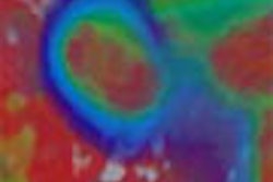
Renal cysts are common and benign; renal carcinoma is not. And distinguishing some high-attenuating cysts from cancer on a typical unenhanced CT scan can be difficult, if not impossible. But researchers from Yale University think they've succeeded in sorting them out fairly accurately, if only to inform the decision for the next exam.
Of course, benign renal cysts are easily characterized at CT when they're well defined, homogeneous, and the same attenuation as water, noted Drs. Ari Jonisch, Ami Rubinowitz, Pradeep Multalik, and Gary Israel from the Yale University School of Medicine in New Haven, CT. "However," they added, "some benign renal cysts may contain high-attenuation fluid (> 20 HU) and are referred to as high-attenuation renal cysts. Unfortunately, at unenhanced CT, these cysts cannot be differentiated from renal cell carcinoma because both may have similar attenuation values" (Radiology, May 2007, Vol. 243:2, pp. 445-450).
The increased attenuation may be secondary to hemorrhage or proteinaceous debris, "and is also dependent on the state of hemorrhage within the cyst," the authors noted.
The similar attenuation to renal carcinoma means that the discovery of a high-attenuating renal mass poses a diagnostic dilemma, requiring additional imaging studies such as ultrasound, MRI, or contrast-enhanced CT to characterize.
The researchers set out to determine if they could characterize such lesions at unenhanced CT by gauging heterogeneity and HU, and found that they could. The retrospective study included 54 pathologically proven renal cell carcinomas in 54 patients and 56 high-attenuation renal cysts in 51 patients.
The 54 renal cell carcinoma patients had been scanned preoperatively with CT, while the 51 high-attenuation renal cyst group had CT findings described as "hemorrhagic cyst," "high-attenuation cyst," or "proteinaceous cyst." The average size was 1.8 cm (range, 1.0-6.1 cm), and patients with dominant polycystic kidney disease or von Hippel-Lindau disease were excluded from the study.
The images were acquired on four -or 16-detector LightSpeed scanners (GE Healthcare, Chalfont St. Giles, U.K.). Scanner collimation settings for the carcinomas varied from 10 mm (n = 1), 7 mm (n = 4), 5 mm (n = 19), 3.75 mm (n = 1), and 2.5 mm (n = 29). Collimation for the renal cysts varied from 10 mm (n = 4), 5 mm (n = 25), and 2.5 mm (n = 27), according to the authors.
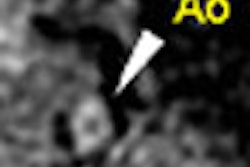
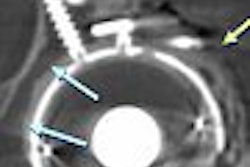
Two readers blinded to the final diagnosis reviewed unenhanced CT images of renal neoplasms and cysts in a random manner. Attenuation was measured at circular or oval regions of interest (ROI) for each mass, they explained, using the largest possible ROI for heterogeneous lesions and avoiding calcified areas.
"The internal heterogeneity of each lesion was also subjectively determined by viewing the lesion with routine abdominal window and level settings and by using a four-point scale," for which 1 was homogeneous and 4 was markedly heterogeneous. Statistical tests included a Bland-Altman regression tree, classification and regression tree, and Shapiro-Wilk normality test, Jonisch and colleagues wrote.
According to the results, the average cyst attenuation for reader 1 was 53.4 HU (range, 23-113 HU) and 38.4 HU (range, 22-60 HU) for reader 2. The average attenuation of neoplasms was lower at 34.7 HU (range, 21-60 HU) for reader 1 and 38.4 HU (range, 22-60 HU) for reader 2. The biggest difference was in heterogeneity. Reader 1 gave the cysts a score of 1 for 55 of the 56 (98%) cysts, while reader 2 gave the cysts a score of 1 for 53 of the 56 (95%) cases.
The carcinomas were far less heterogeneous. Reader 1 gave a score of 1 for neoplasm heterogeneity for 35 of the 54 cases (65%) versus 36 of the 54 (67%) for reader 2.
There was overlap between cysts and tumors in both attenuation and heterogeneity, the authors reported, but no tumor had attenuation greater than 60 HU, while 16 cysts did. Moreover, nearly all cysts (53/56, 95% for reader 1; 55/56, 98% for reader 2) received a score of 1 for heterogeneity.
"Given the distribution of cyst and tumor attenuation values and lesion heterogeneity, a homogeneous mass measuring 70 HU or greater at unenhanced CT has a greater than 99.9% chance of representing a high-attenuation renal cyst," the authors wrote. Knowing what the mass likely represents can be helpful in determining how to evaluate it.
For example, if a homogeneous mass greater than 70 HU were detected in a patient predisposed to develop renal cell carcinoma, ultrasound would be a logical choice for the next exam, rather than CT or MR, which would need to be performed with and without contrast, they wrote. In fact, they note, advances in ultrasound technology in particular have improved its diagnostic power for such lesions.
The authors emphasized that the use of absolute HU alone for the characterization of specific tissue may not be justified, because attenuation can vary widely based on scanner performance, object position within a scanner, change in peak voltage, or scan time. Therefore, assessing the degree of heterogeneity along with attenuation can yield a better idea of what the lesion represents, they explained, though even these results do not provide a definitive diagnosis.
"These findings may be helpful in suggesting the most appropriate next imaging study for definitive characterization," the authors wrote.
By Eric Barnes
AuntMinnie.com staff writer
May 17, 2007
Related Reading
Dobutamine stress echo findings prognostic in chronic kidney disease, April 30, 2007
Group uses electron beam CT for functional CT of kidneys, February 8, 2007
CT gains ground in urology, June 18, 2004
US, MRI challenge CT for classifying renal cell carcinoma, June 17, 2003
Percutaneous radiofrequency ablation may be an option for solid renal masses, April 21, 2003
Copyright © 2007 AuntMinnie.com