The patient cohort was small, but results were nonetheless described as "promising" in a study that assessed the feasibility of using a 256-slice CT scanner for the simultaneous assessment of coronary artery and cardiac function in one-beat, whole-heart studies.
Dr. Akira Kurata of the Ehime University School of Medicine in Matsuyama, Japan, presented his findings last month at the 2006 American Heart Association (AHA) conference in Chicago. The presentation expanded on findings presented earlier this year, at the American College of Cardiology (ACC) show in Atlanta in March.
"In the last few years, CT angiography has been proposed as a noninvasive diagnostic tool for coronary artery disease," Kurata said. "The addition to coronary artery assessment includes myocardial perfusion, cardiac function and analysis, and myocardial ischemia using CT."
The Ehime study included nine patients, three of whom had a history of myocardial infarction and six patients who had experienced angina pectoris. Two of the nine patients also had atrial fibrillation, and seven patients had received invasive coronary angiography.
The imaging procedure began with a test injection of contrast media at 3 to 4 mL/sec (40-60 mL) with saline at 3 mL/sec (20 mL) for optimal scan timing. The scan was obtained without ECG gating and with a single breath-hold. The group used a 256-slice scanner that currently is a work-in-progress (Toshiba Medical Systems, Tokyo).
Because a 256-detector-row CT scanner covers 12.8 cm along the z-axis, one scan covers the whole heart with one 0.5-second rotation. Acquisition time was 1.5 seconds at 0.5-mm slice thickness, achieving a temporal resolution of 250 milliseconds without ECG gating, involving a radiation dose of 2.65 mSv. To further evaluate myocardial enhancement, the scanner can collect perfusion data by acquiring 26 datasets with 256-slice CT at 15-second intervals, Kurata noted, bringing total radiation exposure of 14.2 mSv.
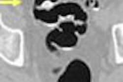
Twenty phases were extracted for coronary imaging and for 2D/3D cine cardiac imaging to assess cardiac function. Coronary arteries also were evaluated, and parameters were obtained for left ventricular function and volume curve, including end-diastolic and end-systolic volumes, as well as ejection fraction calculations. In all nine patients, coronary arteries were assessable without stair-step artifacts.
The analysis found that CT angiography was able to assess 83% (113/136) of coronary segments. Kurata added that some coronary segments could not be assessed due to the small size of the vessels (less than 1.5 mm in 11 cases), calcification in eight cases, and the presence of a coronary stent in four images.
The overall sensitivity, specificity, and positive and negative predictive value for the presence of stenoses of more than 70% (as indicated by coronary angiography) were 100%, 95%, 71%, and 100%, respectively.
Kurata told AHA meeting attendees that while the "number of patients in this study is very small, I hope that 256 CT can expand and study this patient population in the future. In conclusion, 256 CT seems to be a promising, next-generation (technology) for coronary and cardiac imaging, allowing one-beat, whole-heart imaging."
By Wayne Forrest
AuntMinnie.com staff writer
December 7, 2006
Related Reading
Cardiac CT yields significant extracardiac findings, November 26, 2006<
Cardiac CT evolves on multiple fronts, November 20, 2006
CT pioneer test-drives 256-slice scanner, August 21, 2006
Prototype 256-slice CT scanner creates high-res images in a heartbeat, March 14, 2006
Motion-free heart images revealed with 256-slice CT, October 24, 2005
Copyright © 2006 AuntMinnie.com