CHICAGO - Even for indications as serious as suspected stroke, doctors should be aware of the radiation doses being delivered with the multiple CT exams typically delivered during the course of care. The cumulative dose from an initial noncontrast CT scan followed by a CT perfusion study, for example, isn't easy to assess. But a new study from Germany could lead to better dose control and safer scanning.
"We wanted to assess patient radiation exposures to MDCT protocols when assessing patients with acute stroke," said Dr. Matthias Cohnen in a Thursday presentation at the 2006 RSNA meeting. "Obviously, you can do dose estimations using different software solutions (WinDose, ImPact, CT-Expo, etc.), but they won't give you, at the time you need to perform the study, the actual organ doses."
More accurate estimates of local radiation dose during perfusion CT of the brain are needed in light of recent reports of deleterious effects, said Cohnen, who is from Heinrich Heine University and University Hospital Düsseldorf in Germany.
Not mentioned was a rather disturbing photograph from a 2005 Japanese series that has been making the rounds at recent radiology meetings. The photograph depicts a male stroke patient with temporal hair loss -- in a bandage-shaped ring around his head -- following CT and CT perfusion studies (European Radiology, January 2005, Vol. 15:1, pp. 41-46).
According to Cohnen, estimates of local doses due to multidetector-row CT (MDCT), including perfusion studies in addition to cerebral angiography, range from approximately 1-3 Gy in the literature.
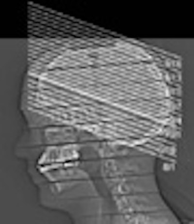
The study measured several standard exam protocols, including sequential CT of the head, CT angiography of the cerebral and cervical vessels, and CT perfusion, simulated using a Somatom Sensation Cardiac 64 MDCT scanner (Siemens Medical Solutions, Erlangen, Germany).
Standard CT of the head employed a typical sequential scanning sequence with collimation of 20 x 1.2 mm, Cohnen said. CT perfusion studies of the brain used a 20 x 1.2-mm detector configuration with tube currents of 200 and 270 mAs, and tube potentials of 80 and 120 kVp. Effective doses were derived from LiF-TLD measurements at the head, the eye lenses, and the thyroid using an Alderson-Rando anthropomorphic phantom.
The results showed effective doses of 1.7 mSv for standard (noncontrast) MDCT of the head, 1.9 mSv for MDCT angiography of the intracranial vessels, and 2.8 mSv for MDCT angiography of the cervical vessels.
 |
| Chart above shows the protocols for the various 64-detector-row MDCT acute stroke protocols; the corresponding radiation doses are below. All data and images courtesy of Dr. Matthias Cohnen. |
 |
The estimated local doses to the brain within the area of the primary beam ranged from 165-445 mGy, Cohnen said. The average estimated organ dose to the brain was estimated at 120-325 mGy. The estimated dose to the eye lenses, though they are not directly in the radiation beam, ranged from 8-35 mGy. The estimated dose to the thyroid ranged from 2-9 mGy.
The results are well-correlated with the literature, he said. Effective doses of combined imaging may range between 4.7 and 9.5 mSv, he said, and of course the particular scanning protocol made a big difference in the actual dose.
"Obviously, you can achieve dose reduction directly proportional when you change your mAs," Cohnen said. "Even more (by a factor of 2.8), you can achieve dose reduction when you decrease your kVp from 120 to 80.... Multidetector technology per se does not mean that you increase your dose compared to single-slice sequential scanners," he said.
By Eric Barnes
AuntMinnie.com staff writer
November 30, 2006
Related Reading
MRI profiles predict utility of reperfusion therapy for stroke, November 22, 2006
Desmoteplase may increase stroke reperfusion window, June 6, 2006
Perfusion CT, MRI agree on most stroke parameters, June 1, 2005
Copyright © 2006 AuntMinnie.com