Radiologists know the value of multiplanar reconstructions (MPRs) for finding what's wrong. In the abdomen, cross-referencing coronal images with the axials can sometimes reveal subtle pathologies that might otherwise be missed.
But some might be surprised to learn that the more detectors rows in the scanner, the more useful MPRs are to the diagnosis.
In a comprehensive examination of abdominal images from several generations of MDCT scanners, researchers from Beth Israel Deaconess Medical Center in Boston discovered that the value of MPRs increased in a statistically significant manner in scanners with the most detector rows.
MRI users take pride in their ability to generate MPRs with every exam, said Dr. Vassilios Raptopoulos in a presentation at the 2005 RSNA meeting in Chicago. But abdominal MPRs can yield major advantages in CT as well, he said. His institution has been using them routinely four years, which is how Beth Israel Deaconess was able to do a study comparing the diagnostic utility of abdominal MPRs in a four-, eight-, 16-, and 64-row scanner.
"Our purpose was to assess the clinically perceived value of routine use of MPR in the interpretation of abdominal CT scans," Raptopoulos said.
The radiologists examined 528 consecutive "mostly gastrointestinal and genitourinary" abdominal and pelvic MDCT scans that were undertaken for various reasons, such as for abdominal pain (acute or chronic), oncology (diagnosis and follow-up), and trauma (acute or postoperative).
The study data included 112 scans from a four-detector machine, 137 from an eight-detector scanner, 148 from a 16-detector scanner, and 131 from a new 64-row machine (Aquilion, Toshiba Medical Systems, Tokyo), Raptopoulos said.
Collimation was set at 2.5-mm for the four-row scanner, 1.0-1.25 mm for the eight- and 16-row machines, and 0.5-0.65 mm for the 64-row scanner. Axial, coronal, and sagittal displays were reconstructed from 5-mm-thick slices, he said.
At the time of clinical interpretation, two reviewers from a staff pool of eight and a resident pool of 15 reviewed the images and subjectively graded the value of MPRs on a five-point scale, with 1 corresponding to no additional value, 2 to subtle value of no clinical importance, 3 to possible value, 4 to important and valuable findings, and 5 to invaluable, abnormality first seen on MPR.
 |
| Sixteen-row MDCT scanner images of a patient with chronic pancreatitis presenting with abdominal pain. MPRs enabled the diagnosis of fibrosing enteritis, and its importance was rated 3. All images courtesy of Dr. Vassilios Raptopoulos. |
 |
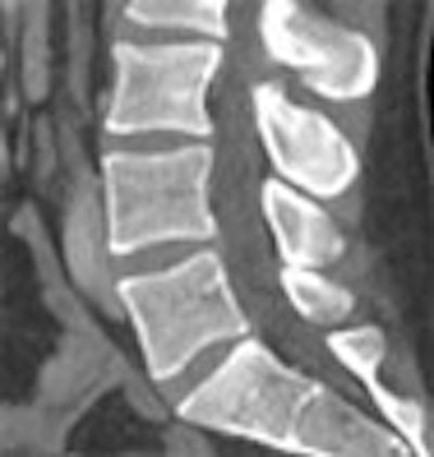
| Sixty-four-row MDCT images of a patient who suffered a moving vehicle accident. Axial images (above) show a very subtle line seen by one observer but missed by a second reader. However, MPRs (below) clearly show vertebral fractures at L1 and L2, adding significantly to the value of the scan -- perceived value of MPR was rated 4. |
 |
According to the results, MPRs were assigned a perceived importance of 1 or 2 in 286 of the 528 patients (54%), 3 in 126 patients (24%), and 4 or 5 in 116 patients (22%). By scanner there was a progressive increase in the perceived value of MPR studies rated 4 or 5 in scanners with more detector rows: 14% of studies in the four-detector scanner, 24% in the eight-detector scanner, 34% in the 16-detector scanner, and 44% in the 64-slice scanner, Raptopoulos said. The improvement was statistically significant only between the 16- and 64-row scanners and between the four- and 16-detector scanners.
 |
| Scans acquired with greater numbers of detector rows were more likely to benefit from the use of MPRs, as expressed in perceived values rated V1 to V4 for the various scanners. Charts courtesy of Dr. Vassilios Raptopoulos. |
 |
"We conclude that the routine use of MPRs is valuable for the clinical interpretation of a large proportion of MDCT studies," Raptopoulos said. The proportion of valuable results increases with the use of higher detector-row scanners."
By Eric Barnes
AuntMinnie.com staff writer
March 22, 2006
Related Reading
Best preps are tailored to VC reading method, November 16, 2005
64 CT slices beat 16 for stenosis and diagnosis of heart disease, November 27, 2005
Modified CT protocols improve orthopedic hardware scanning, October 26, 2005
Scores of detector rows bring opportunities, challenges to CT imaging, April 8, 2005
MDCT yields reliable diagnosis of pancreas divisum, April 14, 2005
Copyright © 2006 AuntMinnie.com


