Was it murder from a blow to the head, or simply a fatal accident? Scientists still don't know how Egyptian pharaoh Tutankhamun, commonly known as King Tut, died.
However, medical imaging is shedding some light on the 3,300-year-old mystery. For the first time, the mummy of King Tut has undergone a CT scan. And based on the results, researchers now believe he may have died from a broken leg that became infected.
King Tut began ruling Egypt around 1355 B.C., and is thought to have died at about age 19 in 1346 B.C., according to Egyptian officials. In 1922 British archaeologist Howard Carter discovered the pharaoh's tomb in the Valley of the Kings, located near what is today Luxor, Egypt. The site was found virtually intact with thousands of artifacts entombed with the mummy of the king.
In 1968 x-rays taken of the remains raised the possibility that King Tut may not have died from natural causes. The films revealed a bone fragment in the skull, leading to the theory that he had been murdered by a blow to the back of the head. But some experts argued that the injury may have occurred postmortem during the mummification process.
Other theories on the cause of King Tut's death range from medical conditions, such as lung disease or a brain tumor, to accidents like a chariot fall or even an insect bite.
But an Egyptian research and conservation project is hoping to lay the murder theory to rest.
Egypt's Supreme Council of Antiquities is conducting a five-year project to learn more about conserving the mummies of Egypt, and also to study health and diseases in ancient Egypt. As part of the project, researchers have begun scanning many of the mummies with computed tomography.
"CT technology enables us to virtually 'unwrap' the mummies without damaging them," said Zahi Hawass, Ph.D., secretary general of the council and head of the research project.
 |
| Inside King Tutankhamun's tomb in the Valley of the Kings, Zahi Hawass, Ph.D. (center), and a team of Egyptian researchers examine the 3,300-year-old mummy as it is removed from its sarcophagus prior to being CT scanned on January 5, 2005. The project is supported by the National Geographic Society and Siemens Medical Solutions. Photo by Kenneth Garrett, copyright 2005 National Geographic Society. |
Hawass' scientific team consists of Egyptian professors of radiology, forensic medicine, and anatomy from Cairo University. The research team also includes a Siemens CT applications specialist and a CT technologist. In addition, three international consultants -- a radiologist and a forensic pathologist from Italy, and an anatomist from Switzerland -- were asked to be a part of the project to review the CT images.
The project is supported by the National Geographic Society and Siemens Medical Solutions of Erlangen, Germany, which donated a Somatom Emotion 6 mobile CT scanner.
The scanner can be transported to wherever the researchers need it. "The system is installed in a trailer so we can do 'house calls' and don't need to transport our patients," Hawass said.
And the system's wide opening allows the mummies to be positioned with minimal movement and disturbance, according to Siemens.
 |
| Hawass (left) looks on as the mummy of King Tut is prepared for CT scanning. Photo by Kenneth Garrett, copyright 2005 National Geographic Society. |
In January, the team removed the mummy of King Tut from its tomb for scanning, which took 15 minutes and produced more than 1,700 images. The Egyptian researchers reviewed the data, and the international consultants confirmed their results, which were released earlier this month.
King Tut's bones indicate that he had a slight build, was well-fed and healthy, and had not suffered from major childhood malnutrition or infectious diseases, the team reported. The king also had a slightly cleft palette and an impacted wisdom tooth, they added.
 |
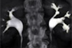
| Dental image taken of the mummy of King Tut. Image courtesy of Egypt's Supreme Council of Antiquities. |
The group was also able to dispel the theory that the young pharaoh was murdered from being struck on the head. "The team found no evidence for a blow to the back of the head, and no other indication of foul play," the researchers stated.
They did, however, find two loose bone fragments in the skull. But the team agreed that the bones could not have been caused by an injury before the king's death, as the fragments would have been stuck in the skull due to the embalming process. The bones matched a fractured cervical vertebra and foramen magnum, which may have been broken during the embalming process, or by Carter and his team after the mummy's discovery, according to the researchers.
 |
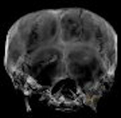
| Image of bone fragment in the skull of King Tut. Image courtesy of Egypt's Supreme Council of Antiquities. |
More surprisingly, the group also discovered a badly broken left femur. The fracture may have punctured the skin and caused a wound infection that killed the young king, according to some members of the team. The CT images revealed ragged edges to the fracture and embalming resin inside the wound -- evidence that supports their belief that the injury occurred while the king was alive.
But of course, chalking up the mysterious king's death to a leg wound would be too easy. Other members of the research team disagreed with the theory, arguing that Carter's team caused the fracture while moving the mummy and that the resin may have been pushed into the fracture while handling the remains. There was no evidence of hemorrhage or hematoma, which should exist if the injury occurred while the king was alive, they added.
Although the exact cause of King Tut's death may never be known, the researchers did rule out murder based on their findings.
"I believe these results will close the case of Tutankhamun, and the king will not need to be examined again," Hawass said. "We should now leave him at rest."
CT images of the mummy of King Tut will be displayed in the National Geographic exhibit "Tutankhamun and the Golden Age of the Pharaohs," along with more than 130 artifacts and treasures from King Tut's tomb, other Valley of the King tombs, and ancient Egyptian sites.
The exhibit will open as part of a four-city U.S. tour on June 16, 2005, at the Los Angeles County Museum of Art (LACMA). The exhibit will also be showing at the Museum of Art in Fort Lauderdale, FL; the Field Museum in Chicago; and the Franklin Institute in Philadelphia. The tour is organized by National Geographic, AEG Exhibitions, and Arts and Exhibitions International, with cooperation from Egypt's Supreme Council of Antiquities.
By Heather Hokenson
AuntMinnie.com staff writer
March 29, 2005
Related Reading
Mikron partners with Field Museum, February 2, 2005
Siemens wraps up mummy imaging deal, January 13, 2005
MDCT solves 400-year-old mystery, November 29, 2004
Mummy dearest: CT helps archaeologists dig deeper, April 23, 2003
Mummyography: Images reveal secrets of ancient remains, December 20, 1999
Copyright © 2005 AuntMinnie.com