Contrast-enhanced ultrasound (CEUS) can allow for reliable quantification of the neovascularization of the carotid artery vasa vasorum, enabling early identification of a condition that can be a precursor to atherosclerosis, according to a presentation at this week's Advances in Contrast Ultrasound: ICUS Bubble Conference in Chicago.
In a comparison of diabetic patients and healthy subjects using contrast-enhanced ultrasound, a multi-institutional research team found that diabetic patients had higher vasa vasorum ratios and, therefore, were at higher risk for cardiovascular disease. They also found a significant association between the presence of HDL cholesterol and lower vasa vasorum ratios, further explaining why "good" cholesterol protects against cardiovascular events.
"For the first time we can analyze the status of the vascular wall of the artery, at the very earliest stage of atherosclerosis," said Dr. Blai Coll of Hospital Arnau de Vilanova in Valenica, Spain.
Coll and Dr. Steven Feinstein of Rush University Medical Center in Chicago will present the group's findings at the Bubble Conference, which will held October 22 and 23 in an educational partnership with the International Contrast Ultrasound Society (ICUS).
Recent research has shown that neovascularization of the carotid vasa vasorum (the blood vessels that supply the walls of the artery) precedes carotid intima-media thickness (IMT) as a surrogate marker for cardiovascular disease. In addition, other studies have found that plaque microvessels originating from the adventitial vasa vasorum show a positive correlation with intimal thickness, indicating that intimal microvessels may play an important role in the development of atherosclerosis, Coll said.
As part of the ongoing quest to improve on the relatively poor performance of current cardiovascular disease screening methods, the study group sought to apply CEUS, which offers better axial resolution than CT and MRI and can yield artery wall information in a noninvasive, reliable, and highly available manner, Coll said.
The researchers studied the carotid vasa vasorum using CEUS in 324 type 2 diabetic patients and a control group of 141 nondiabetic subjects. Participants in the study did not differ in terms of age (59.9 versus 60.2 years old) or gender distribution (61% versus 65.9% male).
After a single operator injected a bolus of 0.5 cc of the Optison microbubble ultrasound contrast agent (GE Healthcare, Chalfont St. Giles, U.K.), cine loops of the carotid artery were recorded. The wall of the common carotid artery on both sides was used as the end-point variable.
A semiautomated software package (AutoIMT, GE) then output the vasa vasorum ratio, which measures the quantity of the contrast microbubbles that are crossing the artery wall (adventitial vasa vasorum/lumen intensity).
 |
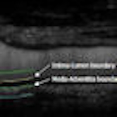
| Carotid artery vasa vasorum ratios are generated by quantifying the amount of contrast that crosses the artery wall. Image courtesy of Dr. Blai Coll. |
Diastolic images were measured using a frame-averaging technique, and clinical examination and laboratory variables were also recorded. The researchers then applied univariate and multivariate statistical analyses.
The researchers found that the diabetic group had a significantly higher vasa vasorum ratio (1.59 versus 0.79, p < 0.0001) than the nondiabetic group.
"That means that the diabetic subjects, even before you get the lumen of an abnormal plaque, presented already [with] a higher neovascularization in the artery wall," he said. "This is the earliest sign of the atherosclerosis in the artery wall."
In other key findings, the researchers found a significant association between HDL cholesterol and vasa vasorum ratio.
"The higher the HDL cholesterol was, the lower was the vasa vasorum ratio," he said. "So it seems that this is a way to explain why HDL cholesterol is protective against cardiovascular events."
The researchers also determined that the nondiabetic group also had significantly higher values of LDL cholesterol and total cholesterol (p < 0.0001). This was likely due to a higher rate of statin use in this group (55% versus 14.9%, p < 0.0001), according to Coll.
From multivariate analyses, the researchers concluded that gender (males were more likely to have higher vasa vasorum ratios), diastolic blood pressure, and the concentration of HDL cholesterol were significantly related to vasa vasorum ratio in both groups of participants.
By Erik L. Ridley
AuntMinnie.com staff writer
October 20, 2009
Related Reading
Contrast US beats MRI, CT for liver lesions, August 17, 2009
Interest grows in noncardiac ultrasound contrast applications, May 21, 2009
CEUS of focal liver lesions in the noncirrhotic liver, May 19, 2009
Contrast ultrasound beats CT, PET in Hodgkin's lymphoma staging, May 6, 2009
New society aims to secure bright future for ultrasound contrast, September 25, 2008
Copyright © 2009 AuntMinnie.com