Researchers in Italy report that low-dose prospectively gated coronary CT angiography (CTA) scans are nearly equivalent to retrospectively gated exams in diagnostic accuracy, while reducing radiation dose by up to 72%, according to an article published in the Journal of the American College of Cardiology.
Also known as step-and-shoot imaging, prospective gating leaves the CT scanner's x-ray beam on for only a short portion of the diastole phase of the cardiac cycle, and turns it off during the rest of the RR cycle. With retrospective gating, the x-ray beam is turned on throughout the RR interval.
The technique was first launched commercially in 2006, and recent clinical studies have validated its effectiveness in reducing radiation dose in coronary CTA exams. Prospectively gated cardiac studies can be performed at doses in the range of approximately 2 to 6 mSv, representing a reduction of 65% to 88% over conventional retrospective gating. Prospective gating does not allow for functional studies of the heart, however.
Informal comparisons of prospective gating's image quality compared to conventional coronary CTA have found the two techniques to be equivalent, but few studies have compared image quality in a randomized patient cohort. Dr. Gianluca Pontone from the University of Milan and colleagues sought to address this shortcoming in a prospective clinical study of 160 patients with a high pretest likelihood of disease (JACC, July 21, 2009, Vol. 54:4, pp. 346-355).
Pontone and his team from the university's cardiovascular services department scanned 160 patients with suspected coronary artery disease, dividing them into two randomly selected groups of patients who were scanned with prospectively gated CT (group 1, n = 78) or retrospective gating (group 2, n = 82), followed by invasive coronary angiography within three days.
All patients with heart rates ≥ 65 bpm were given intravenous metoprolol as a beta-blocker to achieve the target heart rate, and patients were tested for heart rate variability by performing a breath-hold before the exam.
MDCT images were acquired on a 64-detector-row LightSpeed VCT XT scanner (GE Healthcare, Chalfont St Giles, U.K.) using 0.625-mm collimation, 120 kVp, and effective tube current of 700 mAs, following the administration of 90 mL contrast (Iomeron 400 mg/mL, Bracco, Milan) at 5 mL/sec, followed by 50 mL of a saline solution. Bolus tracking was used to initiate the scan.
Prospective electrocardiogram (ECG) triggering (SnapShot Pulse, GE Healthcare) was applied to patients from group 1, with the addition of "padding" of the x-ray window varying from 100 to 400 msec depending on heart rate variability. Patients from group 2 were scanned using retrospective ECG triggering, with ECG pulsing used to determine the x-ray window with a maximum tube current of 700 mAs between 40% and 80% of the RR cycle.
The researchers measured coronary calcium scores using the Agatston method. Group 1 patients were analyzed with vessel analysis software (CardioQ3, GE Healthcare). In group 2, image reconstruction was retrospectively gated to the ECG including x-ray window between 40% and 80% of the RR cycle with a 0.4-mm increment.
Two independent readers blinded to the angiography results read each segment 1.5 mm or larger in diameter, postprocessing the images on a workstation. Any disagreements between readers were resolved by consensus, with significant stenosis defined as ≥ 50% of the coronary lumen.
The group graded image quality for each segment 1.5 mm or larger subjectively as excellent (no artifacts, unrestricted evaluation), good (minor artifacts, good diagnostic quality), adequate (moderate artifacts, acceptable for routine clinical diagnosis), or poor/not evaluable (severe artifacts impairing accurate evaluation), Pontone and colleagues explained.
 |
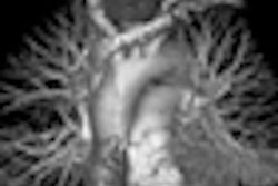
| Above, retrospectively triggered (70% RR) image of a 70-year-old male patient. Below, prospectively triggered (75% RR) coronary CTA image of a 74-year-old male. Images courtesy of Dr. Gianluca Pontone. |
 |
"The overall evaluability of nonstented segments ... was marginally better in group 2 than in group 1 (97% versus 96%, p = 0.05), due to a significantly lower percentage of artifacts (3% versus 8%, p < 0.01)," the group wrote. "Subanalysis of the artifacts showed a higher percentage of blooming artifacts and a lower number of [heart rate-related] motion artifacts in group 2 versus in group 1."
|
Due to the lower number of artifacts in group 2, the evaluability of stented segments was slightly but not significantly better than in group 1 (94% versus 92%), they added.
Accuracy in segment-based analysis was 93% versus 96%, respectively (p < 0.05), including diagnostic segments, and 91% versus 94%, respectively (p < 0.01), including all segments, whereas the accuracy in a patient-based model was 98% in both groups.
The radiation dose was significantly lower in group 1 (5.7 ± 1.5 mSv) compared to group 2 (20.5 ± 4.3 mSv, p < 0.01).
MDCT with both retrospective and prospective ECG triggering delivered a high rate of evaluability (96% for nonstented coronary arteries and 92% for coronary stent evaluation) despite the low-dose protocols. Image quality was considered excellent in 92% of all coronary segments, the team concluded.
Still, despite comparable overall calcium scores between the two groups and a higher number of artifacts in group 1, the prospectively gated scan "seems to present a lower number of artifacts due to blooming effect," they wrote.
And despite similar heart rates between the two patient groups, the higher number of motion artifacts in the prospectively gated group suggests that heart rate variability has a substantial impact on its performance, possibly representing a limitation of the technique.
"Group 2 had a slightly better evaluability and accuracy than group 1 in nonstented segments," Pontone and colleagues wrote. In contrast, prospective triggering did a better job of detecting in-stent restenosis than the retrospective protocol, in a finding that is likely related to technical issues, inasmuch as prospective gating eliminates overlapping slices, thus reducing blooming artifact likely to affect stents and calcified plaques.
Prospective gating generally produces more artifacts in general versus retrospective gating because fewer segments are available for reconstruction, they wrote.
"The prospective ECG triggering allowed a significant reduction of [effective radiation] dose up to 72%," Pontone and colleagues wrote. "Notably, these radiation doses are lower than those of other noninvasive diagnostic tests used in cardiology, including nuclear perfusion scans."
Additional studies are needed to confirm the results, particularly because the study population had a high pretest likelihood of coronary artery disease, a type of cohort for which MDCT results are known to be less accurate compared to low-risk populations.
"Cardiac MDCT with prospective ECG triggering can reduce the radiation exposure with a slight reduction of evaluability and accuracy of noninvasive imaging of coronary arteries and stents," they wrote.
By Eric Barnes
AuntMinnie.com staff writer
July 17, 2009
Related Reading
Repeated calcium scans increase cancer risk, study finds, June 13, 2009
High-pitch cardiothoracic CTA skips breath-holds and high doses, July 7, 2009
Triple-rule-out CT scans benefit from prospective gating, May 28, 2009
Tube current modulation cuts triple rule-out CTA dose, April 15, 2009
JAMA study finds wide variation in cardiac CTA dose, February 3, 2009
Copyright © 2009 AuntMinnie.com