Once upon a time, CT scanners were all pretty similar under the hood, and performance in a given application was predictable across brands. But no longer. Today's state-of-the-art MDCT scanners take widely divergent approaches to difficult imaging targets, such as the heart, and produce different results depending on the application.
Slice wars notwithstanding, there is more to cardiac CT than the number of detector rows. Manufacturers must consider spatial resolution, image noise, artifacts, and temporal resolution when designing their newest CT scanners. Scan speed, coverage area, radiation dose, and cardiac triggering schemes must also be scrutinized -- and corresponding technical trade-offs made.
Endeavoring to outline the basic differences among new CT systems in a nine-minute presentation at Stanford University's International Symposium on Multidetector-Row CT in May was Dr. Mathias Prokop, professor and chairman of radiology at University Medical Center in Utrecht, the Netherlands.
Prokop traced the divergent paths manufacturers have opted for in recent years, comparing their different engineering approaches to the task of imaging the heart, arguably the modality's most challenging and potentially valuable imaging target.
"In the third generation (2004-2005), there was a split in approach between vendors," Prokop said. "Most had chosen a 64-slice design, while Siemens [Siemens Healthcare, Malvern, PA] went back to 32 rows using a z-flying focal spot to double the number of projections," thereby recreating the effect of 64 slices, he said. Prokop called Siemens' dual-energy Somatom Definition, introduced in 2006, the first of the fourth-generation models, delivering 85 msec temporal resolution and the ability to freeze fast heartbeats.
 |
| Siemens' Somatom Definition offers dual-source imaging with two x-ray tubes, z-flying focal spot, and temporal resolution of 85 msec. Image courtesy of Dr. Mathias Prokop. |
For its latest design, the CT750 HD scanner, GE Healthcare of Chalfont St. Giles, U.K., decided to stick with the 64-slice configuration while using a new detector material to boost image resolution and cut image noise, Prokop said. In contrast, Toshiba America Medical Systems of Tustin, CA, and Philips Healthcare of Andover, MA, opted for wide-area detectors to provide greater anatomic coverage.
 |
| Toshiba's 320-slice AquilionOne provides high temporal resolution and 16 cm of anatomic coverage in a single heartbeat. Full organ coverage is an advantage in perfusion imaging of other organs as well. Image courtesy of Dr. Mathias Prokop. |
Toshiba pioneered the area detector scanner with its AquilionOne model, featuring 320 detector rows and 16 cm of coverage to scan the heart in a single beat. Philips sought a compromise with its new iCT scanner among high resolution, speed, and coverage by combining faster gantry rotation speed and a detector that is 8-cm wide, Prokop said.
 |
| Philips' iCT scanner features a wider detector (8 cm), an air-bearing gantry for faster rotation speeds (270 msec), a new x-ray tube with "smart" focal spot, and a 2D grid to reduce the effects of radiation scatter. Image courtesy of Dr. Mathias Prokop. |
Among the four scanners, "detector rows vary from 64 to 320, and the width also varies quite substantially between 1.9 cm and 16 cm, so there's an extreme difference in what the industry decided to do," Prokop said. And while all fourth-generation scanners arrive at a similar place in cardiac imaging, he added, they take very different paths to get there.
 |
| The number of detector rows varies widely between different fourth-generation CT scanners. Image courtesy of Dr. Mathias Prokop. |
There is technical innovation aplenty in the new designs. Philips' air-bearing gantry delivers the fastest rotation speed (270 msec) in the industry, and the 2D scatter grid helps keep scatter radiation from degrading image quality. Toshiba's AquilionOne delivers extremely high temporal resolution, up to 150 msec for whole-heart imaging performed at a single time point, avoiding the need to reconstruct images from several beats and enabling advanced perfusion studies.
 |
| How many heartbeats does each scanner need to cover the heart? Based on detector width, scanners require different numbers of heartbeats to scan an entire heart (14 cm). GE's scanner with prospective gating requires seven beats to image the heart with a 4-cm detector width, including one heartbeat to allow time for table movement. Image courtesy of Dr. Mathias Prokop. |
Two x-ray tubes, along with 3D adaptive filtering, enable Siemens' dual-source scanner to achieve approximately 85 msec temporal resolution that is independent of the patient's heart rate, Prokop said. Meanwhile, GE's new garnet-based scintillator material improves resolution by a third over traditional gadolinium oxysulfide-based materials, while its adaptive statistical iterative reconstruction (ASIR) software reduces radiation dose.
 |
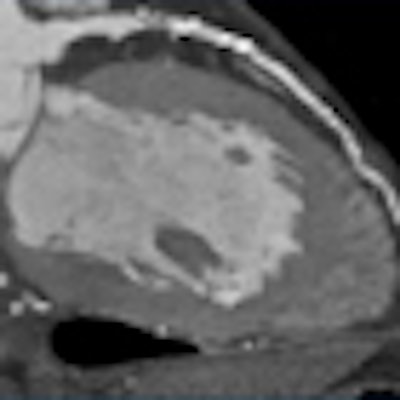
| GE's fourth-generation CT750 HD scanner improves spatial resolution by leveraging the light-handling properties of garnet in its Gemstone detector, as well as ASIR software. Images courtesy of Dr. Mathias Prokop and GE Healthcare. |
 |
| Higher resolution: GE's new scanner delivers spatial resolution of about 230 microns at the 2-mm scan length, versus approximately 300 microns for other 64-slice scanners. |
"Which one is better depends on what you want to do," Prokop said. "If you really want to have the whole heart within one rotation and 16-cm coverage is an issue, you want Toshiba. If you want [prospective triggering], any of the scanners can do that. The [Siemens] dual-source system is the one with 85 msec of temporal resolution -- most are in the range of 140 and 180 msec for prospective triggering. In retrospective mode, of course, you can go down to about 40 to 50 msec," he said, but that resolution is very dependent on heart rate, "so it is not absolutely sure you will get optimal resolution when you do retrospective gating," he said.
 |
| Temporal resolution varies widely with fourth-generation scanners. Siemens' dual-source scanner leads with 85 msec. Image courtesy of Dr. Mathias Prokop. |
To cover the entire heart with cardiac perfusion imaging, the vendors also take different paths, Prokop said. For this emerging technique, Siemens and GE have developed spiral or shuttle mode, respectively, with bidirectional table movement. Wide detectors serve that same purpose at Philips (8 cm) and Toshiba (16 cm); Philips' "jog mode" compensates for its smaller size by switching between two detector views, he said.
The various shuttle and jog modes confer the advantage of greater coverage that is not limited by detector size. However, Prokop said, these techniques are hampered by patient movement and limited temporal resolution, rendering cardiac imaging difficult.
Minimizing radiation dose is a key benefit of prospectively gated CT angiography; however, the technique is no longer vendor-dependent, Prokop said. "It's not a big issue anymore because all of the vendors are focused on creating prospective triggering protocols, and it's extremely important because it allows us to go down in radiation dose substantially compared to earlier approaches," he said.
 |
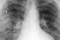
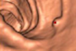
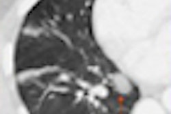
| Crucial factors for CT perfusion are coverage, motion correction, and dose. Above, Toshiba's 320-slice scanner offers whole-organ coverage without the need to reconstruct multiple slices. Images courtesy of Dr. Mathias Prokop and Dr. Patrick Rogalla. |
Advanced CT scanners offer major improvements for perfusion imaging, a promising technique under development for the imaging of coronary vessels, cardiac function, breathing perfusion, perfusion within the brain and myocardium, tumors, and more. However, there are few proven indications to date, and high radiation doses remain an important drawback, Prokop said.
Dual-energy imaging, performed by analyzing the effects of two x-ray signals acquired simultaneously, can offer important new information about tissue properties. Promising applications include stone differentiation, virtual precontrast images, automated bone removal, and iodine maps. Vendors have taken different tacks to enable these applications in their latest scanner models. Siemens uses two x-ray tubes, GE relies on ultrafast kV switching, and Philips uses a split detector configuration, Prokop said.
"We are at a moment where the various scanner [manufacturers] have all decided to take different routes," Prokop said. "They come to very similar results, but you can see there are a couple of applications that favor one scanner over another. Whatever we do we always make compromises, and we hope that within the next generation we will see these approaches come together again -- and we hope that we will see the best of all of them in the generations to come."
By Eric Barnes
AuntMinnie.com staff writer
July 8, 2008
Related Reading
GE focuses on high-res CT with launch of LightSpeed CT750 scanner, May 13, 2008
First 320-slice coronary CTA study shows high image quality, April 7, 2008
Dual-source CT edges into cardiac SPECT turf, March 6, 2008
AquilionOne 320-slice CT scanner spearheads Toshiba RSNA launches, November 26, 2007
Radiation dose slashed in 64-slice coronary CTA, February 15, 2007
Copyright © 2008 AuntMinnie.com