Dr. Daniel Berman has followed the progress of PET for more than three decades, to a point in which today Cedars-Sinai Medical Center in Los Angeles is committed to exploring the modality's efficacy in a wide range of nuclear cardiology applications.
 |
| Dr. Daniel Berman, director of nuclear cardiology/cardiac imaging at Cedars-Sinai Medical Center. |
With a large nuclear cardiology laboratory and multiple cameras, Cedars-Sinai is known for its clinical research, outcome analysis in nuclear cardiology, and software development for the automatic interpretation of cardiac examinations.
Berman is an advocate of PET cardiac imaging with rubidium-82, a radiopharmaceutical that in the past has been somewhat overshadowed by SPECT, in part, because SPECT was a less expensive alternative. He uses CardioGen-82 (rubidium-82, Bracco Diagnostics of Princeton, NJ). In January 2006, the U.S. Centers for Medicare and Medicaid more than tripled reimbursement for rubidium-based myocardial PET perfusion imaging involving multiple studies at rest and/or stress, making the scan more economically feasible for cardiac facilities.
Economic viability
Berman estimates that it would take an average of four or five scans per day to make rubidium-based PET financially viable. Rubidium is delivered by generator once a month for a fee of approximately $30,000. Based on 21 scanning days per month, four patients per day would bring 84 patients to rubidium-based PET. At $350 per dose for the isotope, a facility using the rubidium generator could break even.
Candidates for myocardial perfusion with rubidium-based PET include people who have had, or will likely have, uncertain results with a nuclear scan. A PET scan with rubidium also can be beneficial when imaging a large-breasted woman or woman with implants. PET can overcome artifacts that often are encountered with standard SPECT imaging due to nonuniform soft-tissue attenuation.
 |
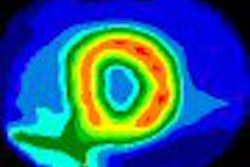
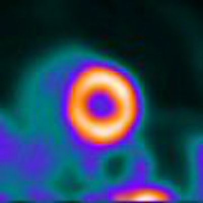
| Above, a cardiac image of a male patient undergoing a SPECT scan. Below, the same male patient's PET scan, in which the results are significantly better with rubidium. All images courtesy of Cedars-Sinai Medical Center. |
 |
"Doctors have learned to live with artifacts and sources of false-positive studies in these women," Berman said. "The PET scans with rubidium, because of the higher energy of the rubidium and the built-in attenuation correction -- that is part and parcel of all cardiac PET studies -- provide the opportunity to have a better imaging test for these women."
The same is true for obese patients, both male and female, who are subject to nonuniform soft-tissue attenuation. Patients who require pharmacological stress because they are unable to exercise also may benefit from rubidium-based PET due to the greater extraction fraction capabilities. In theory, rubidium can take advantage of the very high blood flows achieved with pharmacological stress to identify patients who have mild coronary disease. However, Berman added that this application has yet to be confirmed with research.
Ventricular function
With rubidium-based PET, clinicians also can create an image of ventricular function at the peak of stress, in which a balanced reduction in blood flow may be related to triple vessel or left main coronary artery disease. An abnormality of peak ventricular function might indicate that the disease is present in a circumstance in which it may be underestimated by the perfusion defect, according to Berman.
Cedars-Sinai's nuclear cardiology program can incorporate a 64-slice CT scanner for attenuation correction and coronary calcium scoring of patients. "We are routinely performing coronary calcium scans on all the patients who have PET perfusion scans," he said. "We have found, for example, aortic aneurysms that otherwise were not known to be present."
CT scans on some patients have shown extensive coronary calcification even when a PET scan looks normal. In those cases, Berman said, "we know that the patient must be treated with maximum medical therapy for coronary disease."
Because it is difficult to tell if a patient needs both a CT angiogram and a PET scan, Cedars-Sinai conducts CT angiograms and rubidium-based PET scans at different times.
Continuing research
Cedars-Sinai plans to continue its nuclear cardiology efforts by exploring optimization of its 64-slice CT system and the PET scanner for myocardial perfusion imaging. "The next step will be to help develop and validate quantitative methods to assessing myocardial blood flow rates at rest and at stress," Berman said. "Then we'll optimize ways to take the PET perfusion data and fuse the information with a coronary CT angiogram performed at another time in patients who need both tests."
Cedars-Sinai also will research the efficacy of FDG as an imaging agent for inflamed or vulnerable coronary plaque. Currently, Berman is working with Piotr Slomka, Ph.D., and other colleagues at the facility to image the plaque with PET/CT, using cardiac and respiratory gating with precise registration of the CT and FDG-PET images.
"We think that might lead to better identification of the most vulnerable patients who may be in need of the most aggressive kinds of therapeutic intervention," Berman said.
Cedars-Sinai also is participating in a clinical trial by Lantheus Medical Imaging of North Billerica, MA, for a new PET contrast agent that "appears to have superior imaging characteristics for examining myocardial blood flow," Berman said. If the agent shows promise, he believes that it still will take three or four years before it's available.
By Wayne Forrest
AuntMinnie.com staff writer
April 11, 2008
Related Reading
Study correlates CTA to angiography, myocardial perfusion SPECT, May 25, 2007
Rubidium PET/CT shows potential for coronary artery disease diagnosis, March 30, 2007
Stress MPI with Tc-99m SPECT identifies high-risk obese patients, September 1, 2006
Cardiac SPECT/CT fusion captures SNM's Image of the Year, June 6, 2006
Rubidium-based PET may finally get its due, February 23, 2006
Copyright © 2008 AuntMinnie.com