Low-pitch helical acquisition is typically used in CT cardiac imaging to ensure complete volume coverage and good phase registration over multiple cardiac cycles. The approach is certainly effective, but comes at the price of high radiation doses to patients, and a lack of flexibility in dealing with irregular heart motion. In fact, CTA packs the highest radiation dose of any standard CT application.
A group of researchers has honed in on a potential solution in the form of a "step-and-shoot" acquisition mode for use with 64-slice CT scanners that can reduce even a modulated dose by 50% or more. In a special "hot topic" presentation at the 2005 RSNA meeting, Jiang Hsieh, Ph.D., chief scientist at the applied science laboratory at GE Healthcare (Waukesha, WI), explained the novel protocol and offered his group's preliminary results.
In a standard helical coronary CTA acquisition, the table travels at a constant speed, resulting in much of the volume being scanned more than once and a dose that is far higher than necessary, he said.
"To overcome this (artifact) problem we propose a "step-and-shoot" mode," Hsieh said. "Instead of having the table travel at a constant speed, the table is stationary during the entire acquisition, then we wait until the next (cardiac) cycle and the table moves to the next location for another scan."
The result is very little overlap between the scans and more robust gating, he said. For example, if a patient being imaged in standard helical mode has an irregular heartbeat due to arrhythmia, for example, the radiologist is presented with the dilemma of either keeping the inadequate CT data or discarding it at the price of a data gap between the first and second usable acquisitions.
 |
| Compared to helical mode, the step-and-shoot protocol offers a lower radiation dose, more robust ECG gating, and more flexibility with variable heart rates. All images courtesy of Dr. Jiang Hsieh, GE Healthcare. |
"This can be overcome naturally by the step-and-shoot mode, where you acquire the first cardiac set as before," Hsieh said. "When you move to the next table location and you see there is an irregular heart rate, you stop indexing the table and wait until the next cardiac cycle before you acquire the next set of data."
This function can be further enhanced by the use of advanced ECG gating techniques that base the acquisition on more than the R-R interval peaks. Animal studies have demonstrated the robustness of this technique in handling irregular heart motion, he said.
Interscan delay is a potential problem with the step-and-shoot mode, Hsieh said. Unlike helical mode, in which the table speed is constant, with the step-and-shoot mode the operator must wait until the table settles into each successive location before starting the scan.
The group examined the impact of interscan delay on the total acquisition, using a 16-cm cardiac volume and assuming a relatively long 1.4-second interscan delay and a gantry speed of 0.4 seconds, Hsieh said.
As the graph below shows, the axial scan is very comparable to the helical acquisition using 4-mm detector coverage. However, if detector coverage is reduced to 2 cm or 3 cm as indicated by the curves, total acquisition time increases dramatically, he said.
 |
| Chart depicts analysis of interscan delay between helical and step-and-shoot modes. In the setting of a 1.4-second interscan delay and 0.4-second gantry speed, the scan time increases significantly at a reduced detector coverage of 2 cm or 3 cm. At 4-cm detector coverage, the scan time is comparable to that of helical mode. |
"The longer the acquisition time, the more chance you have to get an irregular heart rate or heart rate variations," Hsieh said.
As for radiation dose, the group compared the step-and-shoot mode to unmodulated or constant tube current acquisition, as well as to 80% dose modulation, wherein mAs is dropped to roughly 20% of the peak value during the fast-moving or undesirable part of the cardiac cycle.
"Compared to both studies, there is a (67% to 83%) dose-savings for no mAs modulation, and even for 80% modulation there is still 52% dose-savings," Hsieh said.
 |
| Step-and-shoot mode cuts the radiation dose vis-a-vis helical MDCT. At 4 cm detector coverage, 1-second interscan delay, 0.4-second scan speed, and 16-cm heart coverage, the average dose saving for non-mAs-modulated scan was 84%; the average dose saving for 80% modulation was 52%. |
The reconstruction algorithm is also crucial for creating high-quality images, he said. Because of the conebeam geometry, regions closer to the x-ray source are much smaller or narrower than regions close to the detector. Most of today's algorithms use extrapolation to approximate the missing samples.
However, this method often leads to conebeam artifacts, Hsieh said. To avoid them, the group applied a conjugate multiscan conebeam reconstruction algorithm for image reconstruction. The so-called FDK algorithm can potentially reduce exposure times due to tighter control of the x-ray exposure window, he explained.
A cardiac phantom was used to test the effectiveness of the proposed reconstruction algorithm on conebeam artifact suppression. The results show that the reconstructed image is visually free of conebeam artifacts, Hsieh said. This has been further confirmed by clinical studies.
"To overcome this (artifact) problem, we make use of the previous and the next scans, and combine the information together to provide complementary information," he explained. "By properly weighing the images reconstructed from both scans, you can really control the amount of extrapolation that occurs."
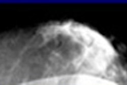
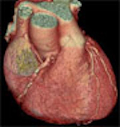
With a 0.35-second gantry rotation speed, the entire heart scan can be completed in less then seven seconds for a wide range of heart rates. Patient studies have confirmed the superior image quality obtained with the proposed method, he said. On each patient, four step-and-shoot half-scans were performed on a GE LightSpeed VCT64 scanner at 64 x 0.625 mm.
 |
| Patient scanned in four step-and-shoot half-scans on a GE LightSpeed VCT64 scanner at 64 x 0.625 mm. |
"We present an alternative acquisition approach to cardiac imaging to take advantage of the large detector coverage enabled by 64-slice scanners," Hsieh concluded. "And we have shown that total acquisition time is comparable to that of helical scan mode, with significantly reduced dose."
Step-and-shoot acquisition combined with the advanced ECG gating and wide detector coverage of VCT enables a significant dose reduction and improved motion artifact suppression in CT cardiac imaging, and the clinical utility of the approach has been clearly demonstrated, he said.
"We have conducted phantom, animal, as well as clinical experiments to demonstrate the efficacy of our approach," Hsieh said.
By Eric Barnes
AuntMinnie.com staff writer
January 16, 2006
Related Reading
Heart rate determines best CTA reconstruction interval, January 2, 2006
Coronary CTA requires dedication, 64-slice minimum, November 30, 2005
64 CT slices beat 16 for stenosis and diagnosis of heart disease, November 27, 2005
Cardiac CT strategies cut dose, keep image quality, November 25, 2005
Motion-free heart images revealed with 256-slice CT, October 24, 2005
Copyright © 2006 AuntMinnie.com