An open-source image processing software package can be a useful tool for evaluating ultrasound images of obese children for signs of fat deposits in the liver, according to research from Brazil published in the May edition of the American Journal of Roentgenology.
Once thought of as primarily an adult disease, fat infiltration of the liver, or hepatic steatosis, is now occurring at rates of 23% to 53% in obese children, and the condition can progress to steatohepatitis or cirrhosis. Although imaging modalities such as ultrasound, CT, and MRI have been used for imaging for this condition, all suffer from a variety of drawbacks.
Ultrasound is economical, widely available, and does not expose children to radiation, and it could potentially be used for hepatic steatosis screening in overweight children to ensure that increases in liver echogenicity caused by other conditions aren't confused with fat infiltration. But the modality is operator-dependent and interpretations are subjective, according to a research team led by Dr. Ricardo Bernardi Soder from Pontifícia Universidade Católica do Rio Grande do Sul (PUCRS) in Porto Alegre, Brazil.
To overcome these drawbacks, Soder and colleagues decided to investigate whether computer-assisted analysis using the ImageJ open-source image analysis software from the U.S. National Institutes of Health (NIH) could help measure liver echogenicity and make ultrasound exams more objective. They also wanted to develop an index of liver echogenicity for both obese and normal-weight children to potentially guide clinicians in the follow-up of children being treated for hepatic steatosis.
In the study, 22 children with an age range of 6 to 11 years were assigned to either obese or normal-weight groups and were paired by sex and age (AJR, May 2009, Vol. 192:5, pp. W201-W205.) All children received an ultrasound exam, anthropometric measurements, and laboratory tests. There were seven boys and four girls in each group.
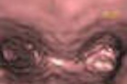
From each exam, an ultrasound image was selected from segment VI of the liver and adjacent liver parenchyma, obtained at the level of the hepatorenal space, according to the authors. The image was digitized as a TIFF file and then analyzed with the ImageJ software, which quantified the echogenicity of the liver parenchyma and right kidney cortex on a scale of 0 to 255 based on grayscale variation. The value for kidney echogenicity was subtracted from the value for liver echogenicity to generate the hepatorenal index.
Although there wasn't a statistically significant difference in the echogenicity values of liver parenchyma in obese and normal-weight children (85.8 ± 26.1 versus 71.5 ± 23.5, p = 0.19), there was a statistically significant difference in the hepatorenal index (33.9 ± 6.6 versus 14.1 ± 6.1, p < 0.001).
In other findings, laboratory tests showed that obese and normal-weight children had statistically significant differences in the values of glucose (p = 0.034), insulin (p = 0.008), triglycerides (p = 0.036), uric acid (p < 0.001), and alkaline phosphatase (p = 0.045).
The researchers acknowledged some limitations of the study, including the unknown number and identity of children with steatosis. Clinical and ethical reasons justified not conducting liver biopsies, however, according to the authors. In addition, the study population was made up of only obese and normal-weight children, and did not include over or underweight children.
Nonetheless, the authors concluded that "computer-assisted analysis of ultrasound liver echogenicity using the ImageJ software is a reproducible and easy-to-use diagnostic tool for calculating the hepatorenal index."
By Erik L. Ridley
AuntMinnie.com staff writer
May 8, 2009
Related Reading
CEUS aids differentiation of small liver lesions, April 3, 2006
Detection of liver metastases with contrast-enhanced ultrasound, March 23, 2006
Contrast-enhanced US useful for differentiating focal liver lesions, January 19, 2006
Contrast-enhanced ultrasound aids in grading liver disease, December 1, 2005
Ultrasound contrast enhances liver imaging, May 11, 2005
Copyright © 2009 AuntMinnie.com