A series of presenters at last week's Society for Imaging Informatics in Medicine (SIIM) meeting in Seattle offered tips on how imaging facilities can maximize the value of their investment in 3D technology. But most of the speakers agreed that 3D's value can be diminished if not managed properly.
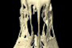
3D images have become so ubiquitous that many surgeons and other clinicians have come to expect 3D as a necessary service from radiologists. Radiology departments run the risk of alienating their referring physicians if they can't produce the types of 3D advanced visualizations that surgeons need, or if the surgeons don't have easy access to 3D images for online review, according to Dr. Gary Wendt, a neuroradiologist at the University of Wisconsin Hospital in Madison.
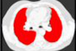
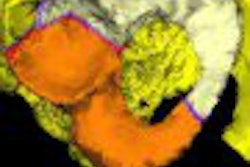
Reinforcing a theme stated throughout the SIIM conference, session presenters emphasized that radiologists must provide added value beyond a traditional written report by adopting and creatively utilizing the various high-tech tools at their disposal -- such as advanced visualization.
"If a radiologist doesn't provide a great product, consumers will find a different solution," Wendt stressed. He told the audience that even if reimbursement for advanced visualization rendering evaporated overnight, his department would continue to provide the service regardless of the added costs it would assume.
The deployment of Service-Oriented Architecture (SOA), a concept for creating and packaging software applications, will make advanced visualization tools easily available for use by other medical specialties. Dr. Keith Dreyer, vice chairman of radiology and director of enterprise imaging at Massachusetts General Hospital in Boston, predicted that SOA will facilitate widespread 3D deployment, and reinforce the need for a 3D lab because of the expertise and efficiency of image production it can deliver.
Properly managed 3D labs bring consistency and high quality to 3D image production, according to Dr. Terri Vrtiska, a radiologist at the Mayo Clinic in Rochester, MN. When individual radiologists handle 3D image reconstruction, there will be variability, she believes.
When the Mayo Clinic established its 3D lab in 2001, surgeons were included from inception in defining the protocols for the types of images they needed. Because of this collaboration, end users rarely need to reconfigure the images, a time-consuming and efficiency-wasting process, according to Vrtiska.
"Three C's -- communication, consistency, and camaraderie -- are the three building blocks which make the operation of a 3D lab successful," according to Vrtiska. Communication among image acquisition technologists, 3D lab technologists, radiologists, and the other clinicians who use images is the key to maximizing the efficiency of the lab and the value and utilization of the images produced.
Advanced visualization workflow is based upon predefined protocols at the Mayo Clinic. The entire protocol for imaging is laid out in a very structured format, and the requirements for 3D imaging are components of this. The result is a consistent output that meets the needs of its constituencies, Vrtiska said.
Camaraderie should never be overlooked, because it's teamwork that makes a 3D lab excel, Vrtiska said. The Mayo Clinic uses a dedicated team of technologists who undergo a six-month training program. Qualifying to become a 3D technologist is a very competitive process, and because of this the technologists are highly self-motivated.
In 2002, the lab produced 100 cases per month. In 2007, it averaged 600 cases per month, and utilization of lab services continues to grow. Turnaround time of one hour or less is achieved, and Vrtiska attributed this to the talent and motivation fueled by camaraderie, communication, and consistency of standards that has been achieved.
By Cynthia Keen
AuntMinnie.com contributing writer
May 20, 2008
Related Reading
3D ultrasound can improve life for sonographers, save money, March 18, 2008
Wise planning helps bring 3D lab to the community practice, February 19, 2008
3D CT planning organizes acetabular fracture fragments into a surgical framework, December 24, 2007
Copyright © 2008 AuntMinnie.com