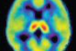
WASHINGTON - With computer-aided detection (CAD) already making its mark in other imaging modalities, researchers at the University of Chicago in Illinois have devised a method that shows how the technology may prove beneficial for tumor detection by PET.
The study, presented at this week's annual meeting of the SNM in Washington, DC, considered 24 consecutive PET scans, which displayed 33 malignant lung tumors in 24 patients. Tumor size, which ranged from 5 mm to 28 mm, with a mean of 15 mm, was determined through the dual-slice CT portion of the PET/CT scan, with biopsies used to prove malignancies.
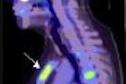
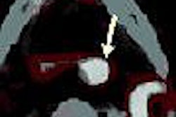
"About two-thirds of these lesions were right up against a normal high SUV structure, which you would think would be a challenge for a CAD algorithm," said Dr. Daniel Applebaum of the University of Chicago, who presented the study results. "Despite this potential problem, (CAD) did fairly well."
The analysis started with a set of 3D volumetric PET images. Each image was interpolated to form an isotropic image with a voxel size of 2.5 mm for the CAD algorithm to be effective.
Tumor enhancement filter
Researchers followed with the application of a selective tumor enhancement filter. Applebaum described the filter as "a mathematical model" to favor tumor characteristics to aid in the detection of initial tumor candidates and suppression of normal anatomic structures.
For each tumor candidate on the original and enhanced image, researchers also determined 12 features, including average standardized uptake value (SUV), standard deviation of the SUV, diameter, spherical characteristics of the tumor, irregularity, and standard deviation for tumor enhancement filter.
"With this initial filter application, our sensitivity was very good -- 94%-- as 31 of 33 tumors were detected," Applebaum said. "As would be expected at this stage, there was a very high false-positive rate -- 21.5% false positives per scan -- which would be an unacceptably high for a screening CAD tool."
FPR correction
To correct the high false-positive rate and increase specificity, researchers used a linear discriminant analysis classifier to distinguish between tumors and false positives. By eliminating the 12 characteristics of a tumor one at a time, researchers returned to assess the performance of the CAD scheme.
"You apply one these features versus another ... and can eliminate the vast majority of false positives," Applebaum said.
The process reduced the false-positive rate from 21.5 per scan to less than one per scan.
The application of the selective tumor enhancement filter resulted in a "very high sensitivity and very high specificity," Applebaum concluded. "We have done it for the thorax and are looking for ways to do it for the rest of the PET scan. There are some different issues when you are outside the lungs, but this seems very promising."
By Wayne Forrest
AuntMinnie.com staff writer
June 5, 2007
Related Reading
Duke researchers combine DR tomosynthesis with 3D CAD, June 4, 2007
Divergent research on CT lung screening sparks more debate, fewer answers, April 19, 2007
CAD performs well in lung nodule detection, February 5, 2007
CAD turns in mixed performance for pulmonary embolism, January 16, 2007
Pilot study: Entropy-driven CAD zips through vast breast image database, August 2, 2006
Copyright © 2007 AuntMinnie.com