A new electronic cleaning method goes beyond simple thresholding-based schemes for distinguishing tagged fluid and fecal material from polyps and folds in virtual colonoscopy. By analyzing the soft-tissue structures associated with submerged polyps and folds rather than just attenuation, the method yields significantly more accurate results with fewer false positives, both with and without CAD.
Dr. Hiro Yoshida discussed his group's results at the 2006 Computer Assisted Radiology and Surgery (CARS) meeting in Osaka, Japan.
Electronic cleansing or digital bowel cleansing, a process for automatically marking and subtracting oral contrast-enhanced residual stool and fluid on CT images, is a key component of efforts to make virtual colonoscopy more accurate and easier to perform.
Traditionally, electronic cleansing has been based on thresholding and gradient analysis of CT image data, a process that gives rise to two image-processing errors. Polyps submerged in tagged fluid have very high CT values, and soft tissues can be completely erased when processed with threshold-based cleansing methods.
In a process called soft-tissue degradation, the pseudoenhancement of polyps caused by surrounding tagged materials causes soft tissues to be removed during electronic cleansing, yielding false-negative readings when applying computer-aided detection (CAD), said Yoshida, who is an associate professor of radiology from Harvard Medical School in Cambridge, MA, and Massachusetts General Hospital in Boston.
 |
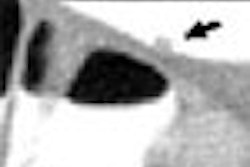
| Illustration of the artifact of degradation of the soft-tissue structures in electronic cleansing method. A thin haustral fold [arrow in (a)] submerged in the tagged materials was erroneously removed after the application of a thresholding-based electronic cleansing method [arrow in (b)]. A small polyp [arrow in (c)] submerged in the tagged materials was erroneously removed by the electronic cleansing method [arrow in (d)]. |
"A second kind of artifact is a false fistula caused by similarity between the lumen tagging boundary and the tagged tissue air layer," he said, wherein the similarity in attenuation values at this interface can produce pseudo polyps and folds due to partial volume effects. "When electronic subtraction is applied you see some kind of false fistula," Yoshida said.
 |
| Illustration of the artifact of false fistula in electronic cleansing. The thin haustral folds sandwiched by the tagged regions and the lumen, called tagging-tissue-air layers (orange arrows, left image), are erroneously removed as the boundaries between lumen and tagged materials (such as the ones indicated by the green arrows), creating false fistulae as indicated by the orange arrows on the right image. |
In the study, principal investigator Wenli Cai with Dr. Michael Zalis; Dr. Janne Näppi, Ph.D.; Gordon Harris, Ph.D.; and Yoshida developed a digital bowel cleansing method that uses a three-step structural analysis process to remove tagged materials without generating spurious objects. In structural analysis, morphologic features are used to distinguish tagged materials from polyps and folds.
"This method consists of three steps: One, analysis of submerged soft-tissue structures in order to remove type 1 artifact," Yoshida said. "Second is an analysis of local isosurface roughness in order to remove type 2 artifact; and third the structural and roughness speed functions are applied to cleanse the tagged regions."
"In the first step, analysis of submerged soft tissues, the method is designed to determine which part of the CAD region should be removed or preserved," he said. "Here the intent is to preserve structures with a ridge-like shape, which is a fold, and cap-like shape, which is a polyp, and all other structures are removed."
These morphologic features are computed from the eigenvalue signatures of a 3D Hessian matrix of a volume I in the CT data. The result is that ridge- and cap-like structures are enhanced while other structures are de-enhanced, and ultimately subtracted from the virtual colonoscopy images, Yoshida explained.
 |
| Submerged soft-tissue analysis was aimed at determining which parts of a tagged region should be removed or preserved. Structures with ridge-like shapes (folds) and cap-like shapes (polyps) are preserved, while other structures are removed. Analysis of local morphologic structures is based on the eigenvalue signatures of the 3D Hessian matrix (H) of a volume (I). All images courtesy of Dr. Hiro Yoshida. |
The second process, isosurface roughness analysis, is aimed at determining whether a voxel is on a thin soft-tissue structure sandwiched between a tagged region and air (tagging-tissue-air layer), or a lumen-tagging (L-T) boundary.
This step is needed because partial volume effect causes significantly overlapping attenuation values around these boundaries, "meaning that the gradient and CT values cannot really differentiate those two structures, unfortunately," Yoshida said.
However, the L-T boundary surface is more irregular than the tagging-tissue-air layer because of the semiliquid nature of tagged fecal materials. As a result, characteristics of the curvedness across scales can differentiate the two types of boundaries by measuring the local isosurface irregularity.
 |
| Example of the structural responses obtained by the analysis of the submerged soft-tissue structures and the local isosurface roughness analysis. (a) Original VC image. (b) The structural response from the Hessian matrix. A submerged fold (orange arrows) is enhanced. (c) The local isosurface roughness. A tagging-tissue-air layer (green arrow) is enhanced. (d) The integration of (b) and (c). Both the submerged fold (orange arrow) and the tagging-tissue-air layer (green arrow) are enhanced, whereas the lumen-tagging layer (red arrow) is suppressed. The integrated structural responses are used in the level set method for preserving the submerged soft-tissue structures while removing the tagged materials. |
As a result of this step, "the surfaces between the lumen and tagging are all deemphasized, and the thin layer between tagging and lumen are preserved or even enhanced," Yoshida said.
The bowel cleansing process works by first calculating responses of the structural enhancement and local roughness at each voxel of the lumen, then initializing the level set front with the soft tissues, and finally, evolving the level set front based on threshold speed function and local roughness, Yoshida explained.
Testing one, two, three
A three-step comparative process was used to evaluate the structure-based analysis scheme.
First an anthropomorphic phantom containing 21 polyps and 11 folds "specifically designed for VC purposes" was scanned twice at multidetector-row CT (MDCT), Yoshida said, once without and then with the addition of tagged fecal material (3.5 mm collimation, 1.6 mm reconstruction interval, 50 mAs, and 140 kVp). The data from the phantom alone was used as a reference standard, while the tagged set was compared to both structure-based cleansing and a previous thresholding-based digital bowel cleansing scheme.
"A nonionic iodine-based contrast agent was used for fecal tagging," Yoshida said. "We analyzed (regions of interest) around the polyps and compared how it was different or the same after electronic cleansing was applied."
In an analysis of 18 ROIs (11 polyps and seven folds), the mean square error of the subtracted ROIs was statistically significantly lower in the new cleansing method compared to threshold-based cleansing.
"A smaller mean square error means that structure-based cleansing results in better cleansing in terms of appearance than (threshold-based) cleansing," Yoshida said.
In the second evaluation, manually cleansed virtual colonoscopy CT data containing polyps 8 mm or larger were used as a reference standard, to which structural analysis and threshold-based cleansing were both compared. (16 cases from Pickhardt et al, New England Journal of Medicine, December 4, 2003, Vol. 329:43, pp. 2191-2200).
In an analysis of 10 ROIs from these cases, including 10 submerged polyps 8 mm or larger, the average mean square error in structure-based cleansing was statistically significantly superior at 2,268 HU² compared to 6,366 HU² for threshold-based cleansing (p = 0.0017).
Finally, the group evaluated the effect of structure-based cleansing on computer-aided detection of polyps. The cohort consisted of 41 patients who underwent low-dose magnesium citrate prep and low-fiber diet with oral administration of 90 mL nonionic iodinated contrast, followed by insufflation and CT scanning in both prone and supine positions, 2.5-mm collimation, 1.25 mm reconstruction interval, 70 mAs, and 140 kVp.
The data were applied to a previously reported CAD scheme that on rigorously cleansed cases yielded 90% by-polyp sensitivity with 1.5 false positives per dataset. When the 41 cases were subjected to threshold-based cleansing, the CAD yielded an average 9.4 false positives per patient, a figure that was reduced by 51% when structure-based cleansing was applied.
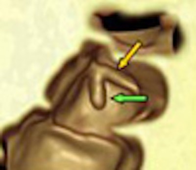
 |
| Example of the cleansing results of a 9-mm polyp submerged in the tagged fecal materials in a clinical VC case by use of a previous threshold-based cleansing (T-cleansing) and a new structure-analysis cleansing (SA-cleansing) methods. (a) 3D volume-rendering view of the polyp after manual cleansing. (b) 3D view of the polyp in (a) after the application of T-cleansing. We observe the degradation of a fold (orange arrow) and a polyp (green arrow) as well as the creation of false fistula (red arrow). (c) 3D view of the polyp in (a) after the application of SA-cleansing. We observer no degradation of the fold (orange arrow) and the polyp (green arrow), as well as no creation of a false fistula. |
"Application on a CAD designed for nontagged (VC) shows that if applied after structure analysis cleansing was done, sensitivity increased from 40% to 80% compared to (threshold-based) cleansing, whereas the false-positive rate is reduced from 9.4 to 4.6 (per case) indicating that the subtraction or cleansing artifacts have been drastically reduced by this method," Yoshida said.
 |
| Example of the cleansing results of a VC case with a large amount of semiliquid fecal materials by use of the new SA-cleansing method. Semiliquid fecal materials having rugged surfaces (violet arrows on the left image) are all removed whereas the submerged folds (orange arrows) are preserved. |
A submerged polyp shown as an example could not be seen in threshold-based cleansing, but was clearly visible after application of the structure-based algorithm, at which point it was visible to CAD as well, Yoshida said.
"A new electronic cleansing method called structure analysis cleansing ... was shown to effectively preserve soft-tissue structures, in which submerged polyps or folds in tagged regions are preserved, and also sandwiched structures such as folds ... are nicely preserved," Yoshida concluded. "The new electronic cleansing method was also shown to substantially improve the performance of CAD, especially to reduce false positives."
By Eric Barnes
AuntMinnie.com staff writer
October 11, 2006
Related Reading
New method corrects for hyperattenuation surrounding tagged VC data, August 8, 2006
Window levels matter when viewing submerged polyps in VC, July 24, 2006
Best preps are tailored to VC reading method, November 16, 2005
CAD struggles through tagged, subtracted VC data, May 18, 2005
Iodine tagging regimen yields best VC results, January 27, 2005
Copyright © 2006 AuntMinnie.com