CHICAGO - Dual-energy CT (DECT) could potentially take urinary stone analysis beyond traditional distinctions of uric acid versus nonuric acid composition, identifying more stone types and enabling greater numbers of patients to be treated noninvasively. However, the presence of imaging artifacts may stand in the way of more detailed analyses in some patients.
Researchers from Harvard Medical School and Massachusetts General Hospital (MGH) in Boston discussed their study of urinary tract stones scanned in phantoms and in vivo with dual-energy CT at the 2008 RSNA meeting.
"Stone composition is a key determinant in deciding treatment strategies for patients with urolithiasis," said Dr. Avinash Kambadakone Ramesh, a research fellow in the department of abdominal imaging and intervention at MGH.
While uric acid stones are usually treated with urinary alkalinization and medical management, nonuric acid stones are treated with interventional procedures such as lithotripsy, ureteroscopy, or percutaneous nephrolithotomy, he said.
"Lithotripsy is also dependent on stone composition, with stones like brushite, cysteine, and calcium oxalate being highly resistant to fragmentation, while struvite stones fragment easily," he explained. If stone composition can be determined with CT, invasive management can often be avoided.
"Dual-source CT has been shown to differentiate between uric acid and nonuric acid stones reliably," Ramesh said.
This study aimed to determine the accuracy of DECT (Somatom Definition, Siemens Healthcare, Malvern, PA) for predicting the chemical composition of urinary tract stones in a phantom model and in patients using dual-source CT.
In the phantom experiment, 65 urinary tract stones extracted from patients (mean, 5 mm; range, 2-18 mm) and of known composition were inserted into kidney-sized potatoes at different locations to simulate stones in the renal collecting system. The stones included 16 uric acid, 10 cysteine, 14 brushite, 12 calcium oxalate, and 13 struvite stones, he said.
The prospective patient study included 18 patients (12 men, six women; mean age, 48 years) with known renal stones (mean, 6 mm; range, 2-24 mm). The patients underwent a routine stone protocol MDCT followed by a focused dual-source CT scan.
After the stones were localized, the phantoms and the patients were scanned with DECT at the same settings: 80 kV and 350-380 mAs for one tube, and 140 kV and 80-98 mAs for the other, at 14 x 1.2 mm and 64 x 0.6 mm, respectively.
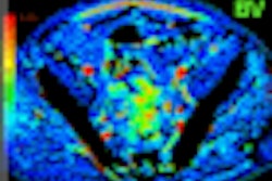
The dual-energy images were then postprocessed using a three-material decomposition algorithm included with the DECT software on the scanner console (syngo VA 11, Siemens). Using the preset algorithm, differentiation between the uric acid and non-uric acid stones was performed, with red representing uric acid stones and blue representing non-uric acid stones.
"The non-uric acid stones were then further differentiated based on density measurements of 80, 120, and 140 kV and by adjusting the slope of the three-material decomposition," Ramesh said. The final determination of stone composition was made by in-vitro analysis of the stones.
Of the 65 stones in the phantom model, 16 were identified as uric acid and 49 as nonuric acid. DECT had a sensitivity and accuracy of 100% in differentiating uric acid and nonuric acid stones in the phantom model -- including stones less than and greater than 3 mm in diameter. Differentiation of cysteine and struvite stones from other stones was possible in 90% (9/10) of the cysteine stones and 84.2% (11/13) of the struvite stones.
A total of 37 stones (mean, 7 mm) were detected in the patient cohort. Seven were identified as uric acid, 23 were identified as nonuric acid, and seven weren't identified as either, using the three-material decomposition algorithm, Ramesh said. That is, the seven stones smaller than 3 mm did not show a red or blue color but remained gray, meaning they could not be differentiated by CT.
Compared to in vitro analysis of stone composition, the diagnostic accuracy at DECT in the patient cohort was 100% for stones 3 mm and larger, zero for the seven stones 3 mm and smaller, and 74% overall in the human cohort, he said.
Further analysis of the nonuric acid stones was made by adjusting the slope of the three-material decomposition, which enabled DECT to distinguish uric acid, struvite, and cysteine stones from other stones in the phantom model, but it did so less successfully in the patient cohort.
The researchers also relied on CT values to distinguish stone types. Uric acid stones had the lowest mean values (228 ± 220 HU), while the calcium oxalate stones had the highest mean values (1,306 ± 220 HU), he said.
In the phantom model, 93% of the cysteine stones and 84% of the struvite stones could be reliably differentiated from the other stones based on the slope of the material decomposition algorithm in addition to CT values, according to Ramesh.
"However, we could not apply this slope method to the patient group because we had not obtained cofirmation of the struvite and cysteine stones from these patients, and also when the slope ratio [was altered], some of the stones showed patchy blue and red colors. Hence, the method was not considered reliable in these patients," Ramesh said.
In the patient cohort, differentiation between uric acid and nonuric acid composition was not reliable for stones less than 3 mm in diameter and in obese patients, he said. But DECT did permit accurate differentiation between uric acid and nonuric acid stones in the phantom study and in most patients.
By Eric Barnes
AuntMinnie.com staff writer
December 4, 2008
Related Reading
CT planning eliminates need for urethrography in prostate, May 12, 2008
New technique may reduce radiation dose with CT urography, September 18, 2007
CT urography helps diagnose cancers, July 4, 2007
Low-dose multidetector CT accurately evaluates urinary stone disease, March 30, 2007
Furosemide beats saline in MDCT urography, September 8, 2006
Copyright © 2008 AuntMinnie.com