CHICAGO - A review of clinical use of breast-specific gamma imaging indicates that the function-based technology changed breast cancer treatment options because additional malignancies were pinpointed by the new modality.
"Gamma imaging can impact treatment management," said Dr. Rachel Brem, a professor of radiology and director of the Breast Imaging and Intervention Center at George Washington University Medical Center in Washington, DC, in her report to RSNA attendees on Wednesday.
"X-rays and magnetic resonance find breast lesions by looking at their shape; breast-specific gamma imaging locates these lesions by how they function," Brem said. She said that gamma imaging has potential as an adjunct to mammography or as a screening tool among high-risk patients.
 |
| Dr. Rachel Brem from the Breast Imaging and Intervention Center. |
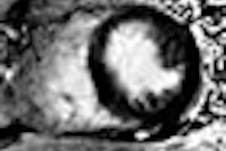
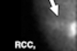
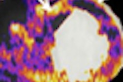
In the study, researchers reviewed 159 scans in which doctors used gamma imaging technology following initial discovery of a tumor. Brem said that in 56 cases, gamma imaging revealed a new suspicious area. Eventually, 45 of those suspicious regions were biopsied, and 14 of those biopsies came back positive for cancer. Overall, that meant that 9% of the women had additional occult cancers, she said.
Nine of the cancers were found in the same breast as the original lesion; five were in the contralateral breast. The cancers that were detected using gamma imaging ranged in size from 0.1 cm to 3.6 cm in diameter.
Brem highlighted one case of a woman who was being considered for lumpectomy, but gamma imaging found another extensive tumor a distance from the original finding, requiring a change in management to mastectomy.
The women in the study were 19 to 93 years of age, with a mean age of 54 years. They all had been diagnosed with one biopsy-proven breast cancer. Approximately 73% of women in the study were classified as having dense breasts, a condition that makes diagnosis with traditional modalities difficult.
The study was funded by the hospital; however, Brem disclosed that she is a board member of Dilon Technologies.
In her discussion of the study, Dr. Mary Mahoney, director of breast imaging at the University of Cincinnati College of Medicine, said that "breast-specific gamma imaging is one of the growing number of molecular imaging modalities that allow us to detect ever-smaller lesions."
She said that Brem's work demonstrated that breast-specific gamma imaging was more than just a tool to find smaller and smaller cancers, in that it was able to guide therapy decisions.
By Edward Susman
AuntMinnie.com contributing writer
December 4, 2008
Related Reading
Dual-head gamma camera increases breast lesion detection, November 28, 2008
Molecular breast imaging offers promise as mammography adjunct, September 15, 2008
Solid-state scintimammography matches breast MRI, July 2, 2008
Breast gamma imaging spots DCIS better than mammo, MR, August 13, 2007
Solid-state dual-head camera supercharges molecular breast imaging, December 19, 2006
Copyright © 2008 AuntMinnie.com