CHICAGO - CT can distinguish the culprit coronary artery lesion in patients with suspected acute coronary syndromes as well as intravascular ultrasound (IVUS) can, according to a study presented on Wednesday at the RSNA meeting.
Intravascular ultrasound is the mainstay imaging modality for the detection and characterization of so-called unstable or vulnerable coronary artery plaques, which are responsible for a significant percentage of myocardial infarcts and strokes.
Distinction of different types of noncalcified plaques, especially thrombi due to plaque rupture from stable noncalcified plaques, has been challenging on CT due to similar attenuation properties. Many researchers have tried and failed to distinguish noncalcified plaques reliably on CT.
But in a cohort of patients who underwent both MDCT angiography and IVUS before coronary intervention procedures, Japanese researchers found that 64-detector-row CT was not only able to differentiate plaque types, but also to identify the culprit lesion in suspected acute coronary syndrome (ACS) patients in every case.
In the study, 33 consecutive stable subjects with anginal chest pain, atypical echocardiogram findings, and suspected ACS underwent emergent coronary CT angiography (CTA) (LightSpeed VCT, GE Healthcare, Chalfont St. Giles, U.K.) just before IVUS, said lead investigator Dr. Iwao Ishibashi from Chiba University Medical Center in Chiba, Japan.
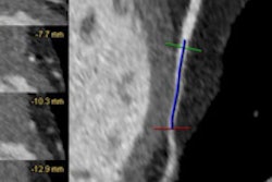
Before coronary intervention, IVUS was performed at 40 MHz using an intrafocus catheter. Images were obtained using a 0.5-mm/sec auto pullback method.
"Plaques were divided into normal plaques; soft [fatty] plaques; and fibrous plaques with or without spotty calcifications, and with or without the presence of remodeling," Ishibashi said in his presentation.
Arterial remodeling was defined as having a remodeling index score of more than 1.05, which is the same as in the study using IVUS, he said.
Patients were excluded if they had evidence of hemodynamic instability, renal dysfunction, history of allergy to iodinated contrast agents, or other contraindications to CTA, Ishibashi said.
The researchers obtained consecutive short-axis images of coronary arteries at CT, and using the major branch as a landmark, confirmed the location and CT values of plaques as measured by two readers and compared with IVUS.
According to the results, 19 patients were ultimately diagnosed as having unstable angina pectoris, and acute myocardial infarction occurred in 14 of those, he said. "Culprit lesions in ACS were diagnosed as thrombus in 19 patients, soft plaque in eight of those, and fibrotic plaque in six of those by IVUS, and the culprit lesions were identified by CT in all subjects," he said.
The mean CT attenuation value of the 19 culprit lesions diagnosed as thrombus by IVUS was 42.8 ± 17 HU, higher than that of soft plaques (n = 8; 35.4 ± 6.4 HU) (p = 0.08). Both were significantly lower than the CT value of fibrotic plaques (n = 6; 83.3 ± 5.7 HU) (p < 0.05), Ishibashi said.
"Spotty calcifications were observed in 50% of lesions in IVUS and 45.5% of lesions in CT," he said. "Regions of spotty calcifications were not significantly different between IVUS and CT."
The ratio of positive arterial remodeling was higher at CT, at 87.1%, compared with 54.8% for IVUS, he said. Plaques with CT values of approximately 50 HU tended to indicate thrombus by IVUS, and most of these patients had creatine phosphokinase elevation at admission. Reproducibility of CT values between the two readers was 0.75.
"In this study, several types of culprit lesions in coronary arteries in subjects with ACS diagnosed by IVUS could also be classified by CT values acquired noninvasively by [MDCT]," Ishibashi said. "CT values in patients with diagnosed thrombi were approximately 40 HU and were significantly higher than those of soft plaques and lower than fibrotic plaques."
CT may be useful to noninvasively differentiate thrombi from certain fibrous plaques in subjects with ACS, he said. And CT was able to detect the presence of spotty calcification and positive arterial remodeling as effectively as IVUS.
MDCT "can evaluate the culprit coronary arteries in subjects with ACS as effectively as IVUS," Ishibashi said. "It may be very useful for noninvasive diagnosis of ACS."
A session moderator said the study was especially interesting for its ability to distinguish fibrous from lipid-rich plaque in culprit lesions. However, because both lesion types have caused problems, there may not be a need to differentiate them, he said.
The moderator also asked if any nonculprit lesions have the same appearance as culprit lesions on CT, and why the soft-plaque CT values were higher than in the literature.
A limitation of the study was that it was based on CT appearance of lesions identified by IVUS, Ishibashi said. He added that if absolute values of plaque types were a little different from the literature, the relative densities of the different plaque types compared to the adventitia in the study were in line with published reports.
Related Reading
By Eric Barnes
AuntMinnie.com staff writer
December 4, 2008
IVUS finds similar antiatherosclerotic effects for two diabetes drugs, November 14, 2008
Coronary artery spasm may trigger acute coronary syndrome, August 15, 2008
64-slice MDCT valuable for assessing coronary artery disease, July 4, 2008
CT angiography outperforms stress testing in diagnosing coronary artery disease, April 16, 2008
MDCT still can't differentiate between noncalcified plaques, January 10, 2006
Copyright © 2008 AuntMinnie.com