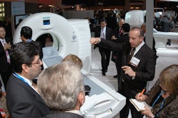
CHICAGO - RSNA 2008 highlights for GE Healthcare include the company's new high-definition CT technology, new digital radiography systems, and new super-premium ultrasound scanner, as well as a product branding initiative that crosses modality lines.
The Chalfont St. Giles, U.K.-based company's branding program calls for the use of three product families that will be applied to systems based on their functionality, rather than their modality. The "Discovery" marque will be used for systems designed for advanced clinical applications, while the "Optima" brand will be employed for systems that target users who want to maximize throughput. The third brand, "Brivo," will be used for economy-oriented products that offer "essential functionality in a straightforward and practical package," the company said.
GE provided an example of the new branding with the launch of two new digital radiography (DR) systems: Optima XR640, a DR system designed for sites that want maximum patient throughput, and Discovery XR650, a DR unit that includes advanced techniques such as the company's VolumeRad digital tomosynthesis package.
Some existing GE products, including the company's recently released high-resolution multislice CT scanner, have been rebranded, with the new scanner shedding the "LightSpeed" prefix long used for GE's multislice CT offerings. That system is now called Discovery CT750 HD.
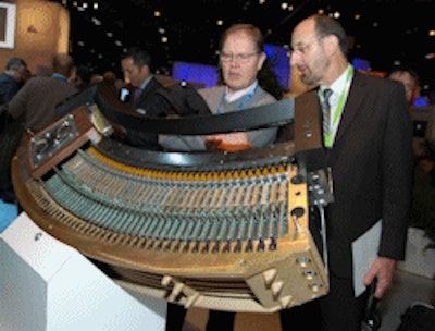
CT
 |
| RSNA attendees examine a detector array from Discovery CT750 HD. |
Dose reduction has emerged as a major theme at this year's RSNA show, and GE is touting the scanner's ability to conduct scans at lower mAs settings with the same image quality as scans conducted at more powerful settings.
A new application, called Gemstone spectral imaging, enhances tissue characterization through its ability to acquire images that separate materials such as calcium, iodine, and water, the company said. The technique also reduces beam-hardening artifacts normally attributed to bone, metal, and iodine and provides tissue contrast optimization.
GE also announced that it is migrating some of the CT750 HD technology to its LightSpeed VCT scanner, creating a new product offering called LightSpeed VCT XTe. GE is making the Extreme HD console available to VCT users, including its iterative reconstruction engine. GE estimates that VCT XTe will be available in late spring 2009, and it will be made available as a console swap for the 3,000 VCT scanners in the field.
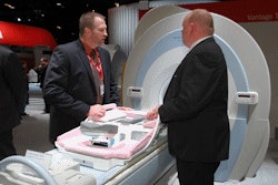
MRI
In the wake of its recently received 510(k) clearance from the U.S. Food and Drug Administration (FDA), GE unveiled its new Discovery MR450 1.5-tesla scanner.
Discovery MR450 features capabilities such as real-time cardiac imaging, single-acquisition multicontrast abdominal images with fat/water separation, and ultra high-resolution musculoskeletal exams.
Built around the company's short-bore, superconducting 1.5-tesla magnet, the system includes a detachable table that can accommodate patients as heavy as 500 lb. The bore has a width of 60 cm.
A newly designed digital receiver extends the scanner's dynamic range by four times compared to the company's previous 1.5-tesla systems, and on-board optical data architecture is isolated from external noise to boost signal-to-noise ratio by up to 27% over conventional nonoptical receivers.
Discovery MR450 also features a newly developed parallel imaging technique, ARC, which was first introduced on the Discovery MR750 3-tesla platform. ARC utilizes a 3D data kernel for enhanced reconstruction and image quality. It can be used for parallel imaging by eliminating errors from patient motion or breath-hold failures and is designed for tight field-of-view prescriptions.
Other new advanced applications include SWAN (T2 star-weighted angiography), which combines 3D T2-based multiecho acquisition with a special reconstruction algorithm. SWAN users can visualize major vessels and large vascular structures and reproducibly image and accurately delineate small vessels and microbleeds. Whole-brain imaging in 3D high resolution can be performed in approximately four minutes.
Also available on Discovery MR750 is Inhance, GE's new suite of Inhance noncontrast-enhanced MR angiography applications. Inhance sequences deliver consistent, reproducible images even in difficult-to-scan anatomies, for fewer rescans and confident differential diagnoses.
Inhance Inflow IR is designed to deliver consistent images of the renal arteries, with an ability to suppress fat, static background tissue, and venous flow.
Also, Inhance 3D Velocity has been optimized to acquire 3D MRA images in the brain and abdomen. This phase-contrast-based sequence enables visualization of complex vascular systems and structures in small detail with enhanced background suppression.
GE plans to begin shipments of its Discovery MR450 in early 2009.
Molecular imaging
In molecular imaging, GE launched Discovery PET/CT 600, which adds to the company's Discovery line of scanners, designed to enable earlier detection and accurate monitoring of disease.
Discovery PET/CT 600 advances include MotionFree technology, using high-sensitivity scintillators, ultra-fast reconstruction enabled by IBM (Armonk, NY) BladeCenter computer hardware, and the high-speed, high-resolution capabilities of the GE BrightSpeed CT.
For clinicians, the Discovery PET/CT 600 features a 70-cm PET and CT field-of-view and a patient table capable of holding patients up to 500 lb. The scanner also offers an increased vertical scan range designed for more flexibility in patient positioning for radiation treatment planning
For administrators, the new scanner offers enhanced start-up time and image reconstruction, allowing for more efficient workflow. Additionally, the IBM BladeCenter offers upgrades in the future. The BladeCenter was designed in collaboration with IBM for this first-ever application in a PET/CT scanner.
Healthcare informatics
On the IT side, GE introduced Centricity PACS-IW Oncology Workflow, a clinical application for Web-based PET/CT functionality.
Available with its newly released PACS-IW version 3.7.1.1, Oncology Workflow brings advanced PET/CT capabilities to a PACS workstation. The latest version of PACS-IW performs automated PET/CT image fusion, reconstruction, and maximum intensity projection (MIP) on the PACS-IW workstation to reduce reading time and increase clinical efficiency.
Users can use multimonitor PACS workstations to review and automatically hang multimodality historical images and simultaneously read fused PET/CT images, as they would read an x-ray, ultrasound, nuclear medicine, MRI, or CT study.
Oncology Workflow also offers new technical achievements. One feature set includes the need to store only PET and CT axial images. When a PET/CT study is launched from the PACS-IW viewer, as well as the viewer from a CD, all of the additional requested orthogonal views are created on-the-fly by the software's advanced image reconstruction algorithms.
Oncology Workflow also offers image manipulation tools, annotations, key images, scanned documents, worklist-driven workflow, comprehensive hanging protocols, referring physician access, and shortcuts.
In addition to Oncology Workflow, version 3.7.1.1 also offers enhanced features for Centricity PACS-IW Breast Imaging Workflow. New features include global shortcuts to increase reading workflow efficiency and a "lights-off" patient folder user interface reducing ambient light, minimizing radiologist eye strain and overall fatigue while reading multimodality breast imaging studies, as well as any other imaging modality on PACS-IW.
GE Healthcare IT also released PACS-IW with international character sets, now supporting French and German languages. These new character sets include support of HL7 integration for connectivity to local HIS, RIS, and electronic medical records.
X-ray
GE is launching two new DR systems at the meeting: Discovery XR650 for users who want advanced clinical applications, and Optima XR640 for those who want to maximize patient throughput.
XR650 includes GE technology such as VolumeRad, a digital tomosynthesis technique that uses a motorized tube head to collect multiple slices of patient data. Tomosynthesis proponents believe it could improve DR's sensitivity in applications such as lung nodule detection. Another technique, dual-energy subtraction, provides a standard postanterior radiograph, a soft-tissue-only image with the bones removed, and an image of the bones highlighting foreign objects and calcified abnormalities, to eliminate bone obstruction from chest or abdominal images.
GE is touting the detective quantum efficiency (DQE) of the system, at 77%. XR650 systems are available in one-, two-, or three-detector configurations and are also available with a portable detector.
In a change for the VolumeRad technique, GE has adapted the technology to support table x-ray shots; previously it was only available on the company's wall stand, for standing patients. The enhancement makes tomosynthesis imaging available for patients who can't stand or who can't be moved, the company said.
As a member of GE's new Optima product family, the Optima XR640 system is designed to maximize patient throughput. The system uses a shared flat-panel digital detector that can be inserted in either a wall stand or table. Features on the unit include automated patient positioning, an automated image stitching tool for long bone and spine studies, and automated tracking between the detector and overhead tube. VolumeRad is not available on XR640.
Both XR650 and XR640 have FDA clearance and will begin shipping in 2009, the company said.
Finally, GE is discussing DR-F, a tube-stand DR system designed for markets in developing countries such as China and India and for rural facilities still using computed radiography. Shipments of DR-F started in September in China.
In the surgical C-arm segment, GE reported that it has shipped 1,200 OEC 9900 C-arms since a consent decree with the FDA was lifted in May 2008. The company said it is planning to begin shipping a cardiovascular version of the product as soon as it receives FDA approval.
Ultrasound
In ultrasound, GE is highlighting Logiq E9, a new super-premium scanner introduced in September. The company is highlighting the system's ability to display ultrasound-acquired images along with images acquired from other modalities, such as CT, MR, and PET. The scanner also includes what GE calls a "GPS-like" technology that enables clinicians to track and mark a patient's anatomy during ultrasound exams.
GE is also announcing its Logiq e Breakthrough 2009 package, which combines real-time clinical images and control capabilities on the same touchscreen user interface on the laptop-based scanner. GE believes the interface helps clinicians focus less on keyboards and controls and more on patients.
In another Logiq e enhancement, GE has added a touchscreen monitor on a boom for use in sterile environments. The configuration has been shipping for several months. On the Logiq 7 scanner, GE features image quality improvements and a new probe, while Logiq 9 will receive an upgrade in the spring. GE is also demonstrating ultrasound computer-aided detection software from Medipattern of Toronto.
Women's health/mammography
In the realm of women's health, GE is demonstrating new capabilities for its Senographe Essential full-field digital mammography system, including the ability to perform biopsies of microcalcifications.
Essential users can now place patients on a portable table to perform prone biopsies, which eliminates the need for a dedicated table. Users also benefit from being able to use a flat-panel digital detector for both imaging and biopsy guidance, as opposed to the charge-coupled device (CCD) digital detector often employed on dedicated biopsy tables.
GE is also highlighting Essential in a mobile configuration, as well as workflow enhancements for mammography. The vendor is planning to offer a mammography viewing workstation, called IDI Mammography Workflow Solution, based on technology acquired through its purchase of German software developer IDI. Essential users will be able to choose either that workstation or the existing Senographe Advantage computer.
Finally, GE is showing digital breast tomosynthesis as a work-in-progress.
By Brian Casey and Wayne Forrest
AuntMinnie.com staff writers
December 2, 2008
Related Reading
GE touts Visipaque study, November 24, 2008
GE launches health IT initiatives, November 18, 2008
GE, UPMC to develop global oncology centers, November 14, 2008
GE debuts women's reproductive assessment software, November 11, 2008
Road to RSNA, Healthcare Informatics, GE Healthcare, November 10, 2008
Copyright © 2008 AuntMinnie.com