CHICAGO - The use of hyperpolarized helium diffusion MRI allows clinicians to assess early changes in the lungs of individuals exposed to secondhand smoke, researchers reported in a presentation on Monday at the RSNA 2007 meeting.
"These findings suggest that breathing secondhand smoke can injure your lungs," said Chengbo Wang, Ph.D., a magnetic resonance physicist in the department of radiology at the Children's Hospital of Philadelphia in Pennsylvania, and formerly of the University of Virginia in Charlottesville.
"We can show that in some people exposure to secondhand smoke extends the time it takes for atoms to travel within lung alveoli," Wang said.
Wang and colleagues at the University of Virginia recruited 60 individuals who were asked to breathe in a small amount of hyperpolarized helium. The patients were placed in a 1.5-tesla MRI unit (Siemens Medical Solutions, Malvern, PA), and lung function images were obtained in about a 30-second period.
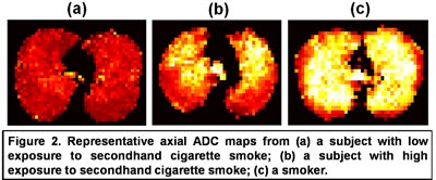
Using software that he developed, Wang then analyzed the distance the atom travels to traverse the lungs. These measurements were translated into scores called apparent diffusion coefficient (ADC) values for each participant in the study.
 |
| Image courtesy of Chengbo Wang, Ph.D. |
Among 23 volunteers who did not smoke and had little exposure to secondhand smoke, about 4% of these volunteers had lung function deterioration.
Among 22 individuals with long-term exposure to secondhand smoke -- living in a household or working where someone regularly smoked for at least 10 year -- about 27% of the subjects exhibited depressed lung function. The difference between the percentage of persons with depressed lung function who were exposed to secondhand smoke and those who were not exposed reached statistical significance (p = 0.047), Wang said.
Among 15 past or active smokers, 67% exhibited depressed lung function, he said. Compared to those not exposed to secondhand smoke, the difference was also statistically significant (p < 0.001).
 |
| Chart courtesy of Chengbo Wang, Ph.D. |
"Helium diffusion MRI detects elevated ADC in subjects with high exposure to secondhand smoke," Wang concluded. "Elevated ADC may reflect lung structural damage or subclinical emphysema. Helium diffusion MRI may be more sensitive than other methods to detect mild emphysematous changes."
However, Dr. Katarzyna Macura, Ph.D., an assistant professor in the department of radiology and radiological science at Johns Hopkins Medical Institutions in Baltimore, urged caution in considering immediate use of the data presented during a press briefing she moderated for the RSNA.
"This is interesting work, but the numbers of subjects is small," she said. She argues that the researchers need to produce evidence that the changes seen on MRI correlate with actual lung damage. She said animal studies depicting this kind of cause and effect are needed.
The study was primarily funded by the Flight Attendant Medical Research Institute in Miami, with additional support from the National Heart, Lung and Blood Institute; the Commonwealth of Virginia Technology Research Fund; and Siemens Medical Solutions.
By Edward Susman
AuntMinnie.com contributing writer
November 26, 2007
Related Reading
MRI depicts small lesions, predicts malignancy in lung cancer, August 3, 2007
Penn researchers utilize new lung MR techniques, March 7, 2007
Workplace cancers cause 200,000 deaths a year: WHO, April 30, 2007
Nonsmoking-related lung cancer more common in women, February 13, 2007
Hyperpolarized helium-3 MRI reveals airflow obstruction in asthma, November 13, 2006
Copyright © 2007 AuntMinnie.com