CHIGAGO - Patients with implanted pacemakers or defibrillators no longer need to be routinely denied much-needed MR scans, according to a presentation Sunday at the 2007 RSNA meeting. Dr. Matteo Quarenghi, Dr. Francesco Sardanelli, and colleagues found that 1.5-tesla MR was safe in their patients with the new generation of cardiac devices.
"The use of permanent implanted pacemakers and defibrillators is widely accepted for the treatment of bradyarrhythmias and tachyarrhythmias. In 2002, 2.4 million patients in the U.S. were implanted with (cardiac) devices," said Quarenghi, whose group is from the University Hospital Policlinico San Donato in Milan, Italy.
The primary concern in these patients is overheating of implanted lead wires because of the currents induced by the radiofrequency fields of the MRI scanner. In pacemakers, heating of the myocardial tissue can result in a failure to pace.
For this study, the presenters enrolled 15 inpatients and outpatients with seven pacemakers and eight implantable cardioverter defibrillators (ICD) that had been implanted between 2001 and 2007. The majority of patients required cardiac MR, followed by abdomen, brain, spine, and breast MR. All scans were done on a 1.5-tesla unit. Cardiac MR studies were done in most cases to evaluate increase in remodeling after myocardial infarction, with the imaging information used to guide surgical restoration, Quarenghi explained.
Before imaging, the group collected data on batteries and lead wires. Therapies such as anti-tachycardia pacing and shocks were disabled, and the devices were checked again after scanning. Markers of myocardial necrosis were sampled before MRI and three hours after MRI.
According to the results, 13 of 15 patients had no significant adverse events during the scans. Two cardiac MR studies were interrupted because of claustrophobia and an adverse reaction to a contrast agent. However, after the MR exam, there were no significant modifications to the 15 devices. Among six inpatients, there was no increase in markers of myocardial necrosis. At six-month follow-up, all 15 patients remained free of device dysfunction.
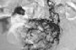
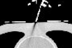
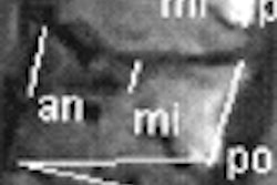
In terms of imaging results, all noncardiac exams were successful. Five cardiac MRIs were also fully realized and three had artifacts, mostly from a "black hole" created by ICD movement, Qaurenghi stated.
Qaurenghi stressed that his group recommends MR scans in patients with devices implanted after 2000, as these tend to have less ferromagnetic components, as well as improved circuitry to protect the device from the magnetic fields.
RSNA session moderator Dr. David Bluemke, Ph.D., noted that the majority of the patients in this study were undergoing cardiac MR exams versus brain or spinal MR. Qaurenghi explained that cardiac MR is his area of expertise so those are the referrals that he gets most often. Bluemke also suggested that turboFLASH imaging could reduce artifacts.
By Shalmali Pal
AuntMinnie.com staff writer
November 25, 2007
Related Reading
MRI of cardiac devices OK, but requires following strict guidelines, January 30, 2007
Radiologists should discuss cardiac implant disposal with patients, January 1, 2007
High-field MRI of the brain safely acquired in pacemaker patients, December 15, 2006
Copyright © 2007 AuntMinnie.com