PHOENIX - In one study it proved very useful; in another it was practically worthless. Now, a third group of researchers has tested the intradecidual sign in obstetric ultrasound, finding overall success in differentiating normal from ectopic pregnancies at an early stage.
Researchers from Harvard Medical School in Boston presented results from the latest study of the intradecidual sign on Monday at the American Institute of Ultrasound in Medicine meeting.
Rapidly differentiating a normal intrauterine pregnancy from an ectopic pregnancy is vital, as the latter is unsustainable and carries the risk of life-threatening hemorrhage for the pregnant patient. Yet despite an increased incidence in recent decades, ectopic pregnancy remains a diagnostic dilemma.
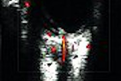
First described in 1986 by Dr. Hsu-Chong Yeh and colleagues, the intradecidual sign of an early normal intrauterine pregnancy is a fluid collection with a slightly echogenic rim (the early gestational sac), located adjacent to the linear echo that marks the uterine cavity.
In contrast, the pseudosac of an ectopic pregnancy is seen on ultrasound as a fluid collection located centrally within the endometrial cavity, said Gloria Chiang, a Harvard medical student who led and presented the latest research.
Yeh's original study found the sign to be 92% sensitive and 100% specific in ruling out ectopic pregnancy among 41 patients. He and his co-authors deemed the sign useful for diagnosing intrauterine pregnancy at an early gestational age ranging from 25 days to five weeks (Radiology, November 1986, Vol.161:2, pp. 463-467).
But a subsequent 1997 study of 102 patients led by Dr. Faye Laing at Boston's Brigham and Women's Hospital achieved very different results.
In that study, the intradecidual sign achieved sensitivity of only 34% to 66%, and specificity of 55% to 73%. The four reviewers in that study also miscategorized three to five ectopic pregnancies as demonstrating the intradecidual sign.
"We decided to repeat the study using a retrospective study design," Chiang said in her AIUM presentation. Chiang's co-authors include her advisor Dr. Deborah Levine, director of obstetric and gynecologic ultrasound at Beth Israel Deaconess Medical Center (BIDMC) in Boston.
Out of the more than 16,000 pelvic and obstetric ultrasounds performed at BIDMC from 1999 through 2001, the Harvard researchers identified 187 exams in which transvaginal sonography revealed an intrauterine fluid collection without a yolk sac or embryo.
They then selected four significant images from each case for a blinded retrospective review by three radiologists: one experienced in obstetric radiology, one abdominal specialist, and a women's imaging fellow.
The reviewers graded the presence of the intradecidual sign on a scale of 1 to 5, with 1 as definitely absent and 5 as definitely present.
"Of the 34 ectopic pregnancies, all of them had a median score of 1 or 2, meaning that the sign was definitely or probably absent," Chiang reported. "Most of the normal intrauterine pregnancies had a median score of 4 or 5, meaning that the sign is definitely or probably present."
Including all three reviewers, the sign's sensitivity was 60%-68%, and specificity was 97%-100%. Both the abdominal radiologist and the women's imaging fellow had one false-positive, where they called an intradecidual sign in an image that was actually an ectopic pregnancy.
"One thing we want to emphasize in our study is it's important to see the unchanging appearance of the sign on multiple views," said Chiang, showing a false-positive case where only the transverse view showed a ring-like structure.
The latest research also found that the intradecidual sign's accuracy increases when the hCG (human chorionic gonadotropin hormone) values are 2,000 or greater, or the mean sac diameter is 3 mm or larger.
"Although we didn't get a sensitivity or specificity as high as originally reported by Yeh, we did find the intradecidual sign is useful in ruling out ectopic pregnancies," Chiang said. "And our statistics were higher than those reported in Laing, et al's study."
What would explain the difference? One possibility was that the unchanging appearance of the sign on different views could not be confirmed for all cases in the Laing study, where reviewers sometimes had only two images for diagnosis.
The evolution in ultrasound equipment could also have boosted performance in recent years, suggested Chiang. Her group's findings are slated for publication in an upcoming edition of the American Journal of Roentgenology.
By Tracie L. ThompsonAuntMinnie.com staff writer
June 22, 2004
Related Reading
The Developing Fetus: First and Second Trimesters, March 16, 2004
US distinguishes between ectopic pregnancy, corpus luteum, February 9, 2004
All signs point to ectopic pregnancy on ultrasound, October 10, 2001
Copyright © 2004 AuntMinnie.com