Does virtual colonoscopy miss dangerous polyps? Not necessarily, say researchers from NYU Medical Center in New York City, based on a preliminary study. VC found most larger polyps (missing many smaller ones), while conventional colonoscopy missed a few of all sizes.
The researchers concluded that both screening exams are probably adequate if the appropriate follow-up interval can be determined and adhered to. Perhaps more important, they dared to make specific follow-up screening recommendations for virtual colonoscopy findings, a subject of keen interest to VC providers as they seek to establish reliable, evidence-based practice standards for the emerging modality.
"A major concern is that CT colonography (VC) performs poorly for detecting small polyps," wrote authors Dr. Michael Macari, Dr. Edmund Bini, Dr. Stacy Jacobs, and colleagues from the New York University Medical Center. "However, many small colonic protrusions are normal colonic mucosa or hyperplastic polyps at pathologic evaluation. These lesions have no clinical potential to become carcinoma. Although small adenomas are not uncommon, their clinical significance has been questioned" (American Journal of Roentgenology, July 2004, Vol. 183:1, pp. 127-134).
The adenoma-carcinoma sequence -- the process by which most colorectal carcinomas are thought to develop from precursor adenomas (though most precursor adenomas do not progress to malignancy) -- is generally estimated to be quite long, up to 10 years. For this reason, many researchers believe that colorectal cancer screening at prescribed intervals should be able to detect most lesions before they reach malignancy.
As part of the NYU group's effort to determine the optimal screening algorithm, the researchers sought to determine the histology and clinical significance of small polyps missed at VC compared to conventional colonoscopy, and determine the appropriate algorithm for follow-up screening, surveillance, and intervention.
The study examined 186 men (ages 40-87 years, mean 62.3) from a Veteran Affairs Medical Center in Manhattan, all with symptoms such as a change in bowel habits, or histories that put them at above-average risk of colorectal polyps or cancer. The subjects underwent same-day virtual colonoscopy, followed immediately by conventional colonoscopy. Each polyp detected with conventional endoscopy was "measured, photographed, biopsied, and histologically analyzed," the authors wrote, and endoscopy results were compared to those of virtual colonoscopy.
A day before the study, the subjects underwent purgative bowel cleansing with two 45-mL doses of Phospho-soda (Fleet Pharmaceuticals, Lynchburg, VA) or 4 L of polyethylene glycol (Braintree Laboratories, Braintree, MA). The colon was manually insufflated with room air prior to CT imaging.
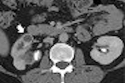
The virtual colonoscopy images were acquired on a Plus 4 Volume Zoom multidetector-row CT scanner (Siemens Medical Solutions, Malvern, PA) using a 4 x 1-mm detector configuration, 120 kVp, 0.56-sec. rotation, 50 effective mAs, and a 30-second breath-hold. Images were reconstructed in 1.25-mm-thick sections using a reconstruction interval of 1 mm, and reviewed on a Vitrea 2 workstation (Vital Images, Plymouth, MN) by a single radiologist experienced in VC interpretation.
Conventional colonoscopy was performed by a board-certified gastroenterologist or senior fellow under direct supervision of the gastroenterologist, and histologic analysis was performed in every case in which there was a biopsy.
Results
In all, colonoscopy found 191 polyps in 97 patients, 53 of which were prospectively identified on virtual colonoscopy -- an overall sensitivity of just 27.7% for virtual colonoscopy (95% CI, 22.1% to 35.6%). VC failed to detect most of the smallest lesions, 5 mm or under, of which the modality found just 21/143 (15%). Virtual colonoscopy detected 12/26 (46%) lesions between 6-9 mm (95% CI, 26.2% to 67.2%), and 20/22 (91%) lesions 10 mm and larger (95% CI, 70.9% to 99.1%).
Of the 122 lesions 5 mm and smaller not detected with VC, 30 were normal colonic mucosa, 37 were hyperplastic polyps, 43 were tubular adenomas, seven were villous adenomas, and four were of unknown pathology, according to the results. In all, 41.9% of these lesions were adenomas.
"Histologic analysis of the 14 lesions measuring 6-9 mm that were not detected on CT colonography showed that one was colonic mucosa, three were hyperplastic polyps, seven were tubular adenomas, one was a tubulovillous adenoma, and two were unknown pathology," the authors wrote. Of these, 57.2% were adenomas.
For its part, conventional colonoscopy was seen as an imperfect gold standard. Eighty-nine patients showed no abnormality with colonoscopy compared to 74 on VC, for an overall patient specificity of 83.1%. The smaller lesions detected with VC but not with colonoscopy were written off as false-positives, including 17 (60.7%) 5 mm or smaller, and eight (28.6%) 6-9 mm lesions. Three polyps detected on VC but not colonoscopy (10.7%) were 10 mm and larger, so follow-up conventional colonoscopy was performed in these three patients. These exams revealed an area of severe diverticular disease, a 10-mm tubulovillous adenoma, and a 17-mm tubular adenoma.
"When considering what constitutes a clinically significant polyp, three factors are of special importance: histology, gross morphology, and size," the authors wrote. "Polyps that are flat, sessile, or irregular, especially lesions that are ulcerated, tend to have a more advanced histology, such as tubular or villous adenomas. Polyps that are pedunculated tend to have a more benign histology, and include hyperplastic and inflammatory polyps and tubular adenomas However, gross morphology alone cannot be used to determine clinical significance because some sessile lesions are hyperplastic and some pedunculated lesions contain villous histology."
Size, while an imperfect predictor of morphology, has turned out to be the simplest and most useful indicator of polyp abnormality, the authors wrote, citing several studies. In one, 657/1228 polyps smaller than 10 mm were adenomas, but neither of the two carcinomas found was smaller than 5 mm (European Journal of Surgery, October 2001, Vol. 167:10, pp. 777-81).
In another study, polyp size was the most important factor in determining malignancy; no invasive carcinoma was found among 5,027 small adenomas 5 mm or smaller (International Journal of Colorectal Disease, 1997, Vol. 12:5, pp. 267-271).
Most colorectal cancers do follow the slow adenoma-carcinoma sequence, and most diminutive polyps never progress to carcinoma, the authors noted. As a result, the eventual goal is to create a rescreening algorithm frequent enough to catch a high percentage of advanced adenomas before they become cancerous, but remaining infrequent enough to be cost-effective while facilitating compliance. Current American Gastroenterological Association guidelines call for screening with sigmoidoscopy or double-contrast barium enema every five years, they wrote.
As for VC findings, the authors recommended against routine conventional colonoscopy follow-up if a lesion 5 mm or smaller is found.
"Many of these lesions will not be adenomas but hyperplastic polyps, normal colonic protrusions, or retained adherent fecal material," they wrote. If VC finds a lesion 6 mm or larger, colonoscopic follow-up is recommended. However, "if the patient has severe cardiac or pulmonary dysfunction, has an allergy to sedatives, has a bleeding diathesis, or is of advancing age, short-interval follow-up every one to two years may be warranted instead of polypectomy," they stated.
Another recent study recommended an 8-mm size cutoff for colonoscopic follow-up, they noted, commenting that this study's approach may be cost-effective as long as inherent variability in measuring polyp size is considered.
A principal limitation of the study included the lack of flat adenomas in the study cohort. Flat adenomas tend to be aggressive and are difficult to detect on virtual colonoscopy. Follow-up studies over several years will be needed to verify the study recommendations. And the survey did not address the issue of cost-effectiveness, which will be a critical factor in determining the appropriate cutoff size of VC findings referred for colonoscopy, the authors noted.
"CT colonography detects most large polyps, but performs less well for smaller lesions," they concluded. "Because of the natural history of colorectal cancer and the adenoma-carcinoma sequence, missing these small lesions may not be clinically important if an appropriate screening interval is established. We propose follow-up in five years if (VC) findings are normal, follow-up in three to five years if a diminutive lesion is detected, and conventional colonoscopy if a lesion measuring 6 mm or greater is detected."
By Eric Barnes
AuntMinnie.com staff writer
August 18, 2004
Related Reading
VC and gastroenterology: friends in need?, July 16, 2004
Group credits 3-D reading for best-ever VC results, October 15, 2003
Workflow issues key in virtual colonoscopy, January 15, 2003
Multiobserver performance trial assesses utility of screening VC, November 27, 2002
Copyright © 2004 AuntMinnie.com